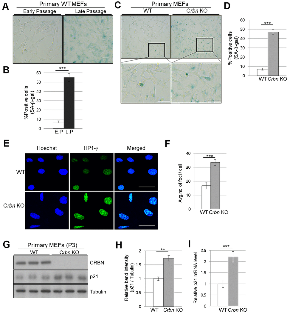
Figure 3.The absence of CRBN induces the markers of cellular senescence in the primary mouse embryonic fibroblast. (A, C) Representative staining images showing SA-β-Gal(blue-stained cells) in primary MEFs. E.P = early passage (P3), L.P = late passage (P8-10) Scale bar = 100μm. (B, D) Quantification of SA-β-Gal-positive cells shown in (A, C) respectively. Results are expressed as the percentage of stained cells (mean ± SEM). (E) Representative images for HP1-γ foci by immunofluorescence staining. (F) Quantitative analysis of HP1-γ foci per cell. (G) Endogenous levels proteins as determined by western blot analysis using extracts from the WT and CRBN KO MEFs. The passage numbers were indicated in the figure. Proteins were subjected to immunoblotting with the anti–CRBN, anti-p21, and anti–Tubulin antibodies. Tubulin was used as a loading control. (H) The relative band ratio of CRBN and p21 to tubulin as determined by densitometric analysis of the blots in (G). (I) Total RNA was isolated from each type of MEFs and subjected to qRT-PCR to measure the mRNA expression of p21. Expression was normalized against β-actin mRNA levels. Fold changes in the mRNA levels relative to control WT MEF is shown. The results shown are representative of five independent experiments. *P < 0.05; **P < 0.01; ***P < 0.005; n.s., not significant.