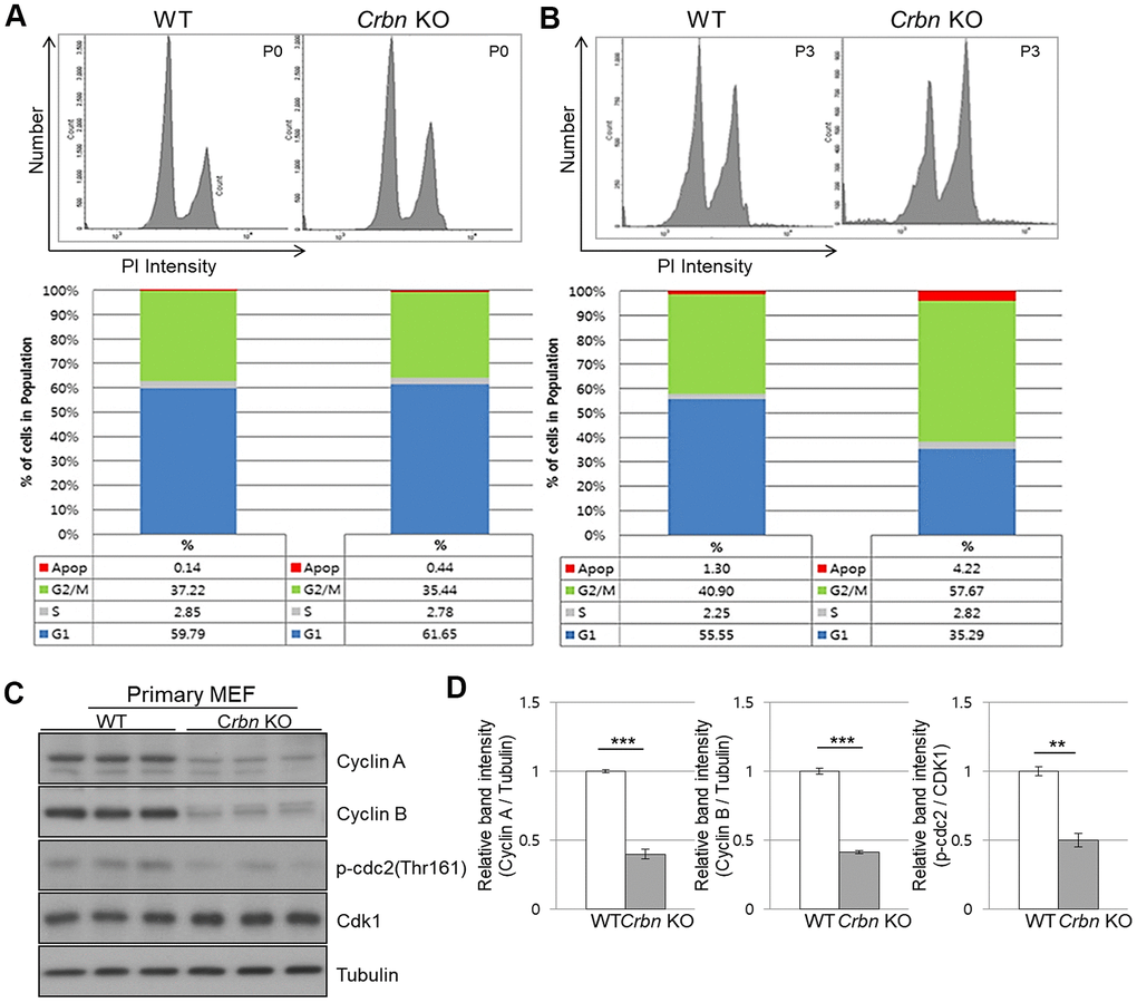
Figure 4.CRBN deficient fibroblast exhibits G2/M cell cycle arrest. (A, B) PI staining and cell cycle distribution analysis of WT and CRBN KO Primary MEFs in P0 and P3. Cells were stained with PI and analyzed for cell cycle distribution using flow cytometry. Representative images of flow cytometry plots are shown. The graph indicates the distribution of each cell cycle phase with different colors, G0/G1(blue), S(gray), and G2/M(green) phases. (C) Western blots analysis using extracts of MEF cells in the early passage were immunoblotted with the anti-Cyclin A, anti-Cyclin B, anti-Cdk1, anti-p-cdc2, and anti–Tubulin antibodies. Tubulin was used to confirm equal protein loading. (D) Relative band intensities as determined by densitometric analysis of the blots in (C). The results shown are representative of five independent experiments. *P < 0.05; **P < 0.01; ***P < 0.005; n.s., not significant.