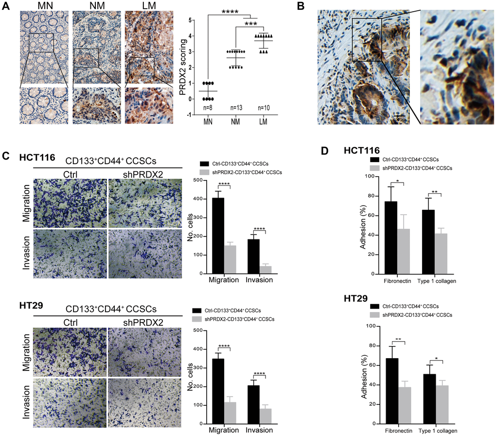
Figure 4.PRDX2 depletion decreases the migration and invasion capacities of CCSCs. (A) Left panel: Representative IHC staining for PRDX2 expression in matched normal (MN) colon tissues, nonmetastatic (NM) colon cancer tissues and colon cancer tissues with live metastasis (LM). The corresponding high-magnification images are also shown. Right panel: The PRDX2 expression intensity scores for 25 patient tissues, including MN (n = 8), NM (n = 13), and LM (n = 10). Statistical analysis: Fisher’s exact test, ***p < 0.001, and ***p < 0.0001. (B) Representative IHC staining for PRDX2 in the invasive front reveals clustering of tumor cells with PRDX2 accumulation. (C) Invasive and migratory capacities of CD133+CD44+ CCSCs generated from HCT116/HT29 control or shPRDX2 cells (left panel). The bars represent the means ± SD of invaded/migrated cells from three independent experiments performed in duplicate (right panel). Statistical analysis: Student’s t-test, ***p < 0.001, ****p < 0.0001. (D) Adhesive capacity of HCT116/HT29-control- or -shPRDX2- CD133+CD44+ CCSCs to fibronectin and type 1 collagen, respectively. Percent adhesion was calculated as the number of adhesive cells/adhesive cells + nonadhesive cells. The data are presented as the percent of adhesive cells observed in three fields per assay and are expressed as an average of triplicate determinations. Statistical analysis: Student’s t-test, *p < 0.05, and **p < 0.01.