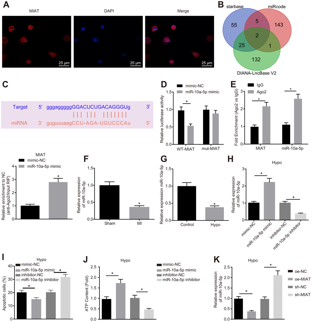
Figure 2.MIAT downregulated miR-10a-5p and suppressed cardiomyocyte apoptosis. (A) The location of MIAT in cardiomyocytes assessed by FISH assay (400 ×). (B) The downstream miRNAs of MIAT predicted by Starbase, miRcode, and DIANA-LncBase V2. (C) The predicted binding sites between MIAT and miR-10a-5p. (D) The interaction between MIAT and miR-10a-5p detected by dual-luciferase reporter assay. (E) Binding of MIAT to miR-10a-5p relative to IgG confirmed by RIP assay. (F) miR-10a-5p expression normalized to U6 in the myocardial tissues of mice assessed by RT-qPCR (n = 10). (G) miR-10a-5p expression normalized to U6 in the hypoxic cardiomyocytes assessed by RT-qPCR. (H) miR-10a-5p expression normalized to U6 in the hypoxic cardiomyocytes after transfection with miR-10a-5p mimic or miR-10a-5p inhibitor assessed by RT-qPCR. (I) Apoptosis of hypoxic cardiomyocyte after transfection with miR-10a-5p mimic or miR-10a-5p inhibitor analyzed by flow cytometry. (J) The content of ATP in the hypoxic cardiomyocytes after transfection with miR-10a-5p mimic or miR-10a-5p inhibitor. (K) miR-10a-5p expression normalized to U6 in HL-1 cells under hypoxic conditions after alteration of MIAT determined by RT-qPCR. * p < 0.05. The above data were all measurement data, and expressed as mean ± standard deviation. The unpaired t test was used for comparison between two groups, and one-way ANOVA was applied for comparison between multiple groups followed by Tukey’s post hoc test. All the data was collected from 3 independent experiments respectively.