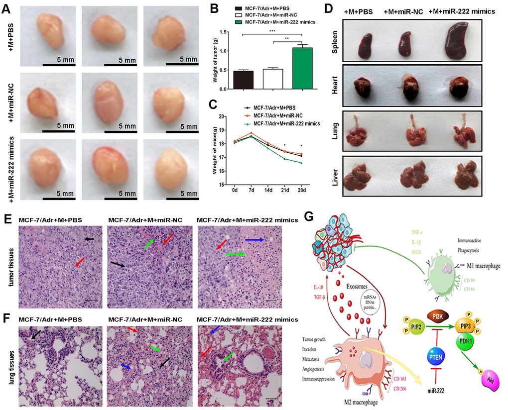
Figure 5.Exosomal miR-222 stimulate tumor growth via macrophages M2 polarization and pre-metastatic niche formation in vivo. (A) MCF-7/Adr mixed with macrophages transfected with miR-222 mimics or miR-NC were implanted subcutaneously into mice, and transplanted tumors and tissue samples were collected 4 weeks later. Representative images of subcutaneous tumors were shown (bar indicates 5 mm). (B) Weight of tumor was measured. (C) Weight of mice was monitored weekly. (D) Appearance of tissue samples including spleen, heart, lung, and liver were shown. (E) Representative HE staining images (magnification × 200) of tumor tissues showing the fibrous tissue separation (black arrow), malignant cell proliferation (red arrow), necrosis area (green arrow), and macrophages infiltration (blue arrow). (F) Representative HE staining images (magnification × 200) of lung tissues showing the macrophages infiltration (black arrow), defuse hemorrhage (red arrow), stromal cells necrosis (blue arrow), and bronchus exudation (green arrow). (G) Schematic model of exosomal miR-222 within A/exo promoting macrophages M2 polarization and breast cancer metastasis. Data are shown as mean ± SD, n = 3 independent experiments; * P<0.05, ** P<0.01, and *** P<0.001 compared with controls.