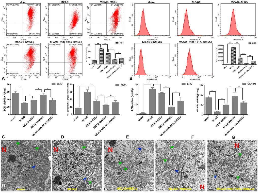
Figure 9.IhOM-MSCs transplantation enhances the mitochondrial function recovery after ischemic stroke. (A, B) Mitochondrial membrane potential was measured for using the JC-1-labeled by flow cytometry. (B) Levels of ROS were evaluated by flow cytometry. (C–F) To measure oxidative stress levels in cells, MDA/LPO contents and SOD/GSH-Px viabilities were measured by biochemical kit respective. (G) The mitochondrial and endoplasmic reticulum morphometric ultrastructural analyses were observed by transmission electron microscopy (TEM) in neurons of penumbra cortex. As shown in the figure: the white dotted lines represent the outlines of the nuclei, letter N represent nucleus, green arrows represent mitochondria and blue arrow represent endoplasmic reticulum. (OM-MSCs were replaced by MSCs in the figure. All data are presented as the mean value ± SD. *p<0.05, **p<0.01).