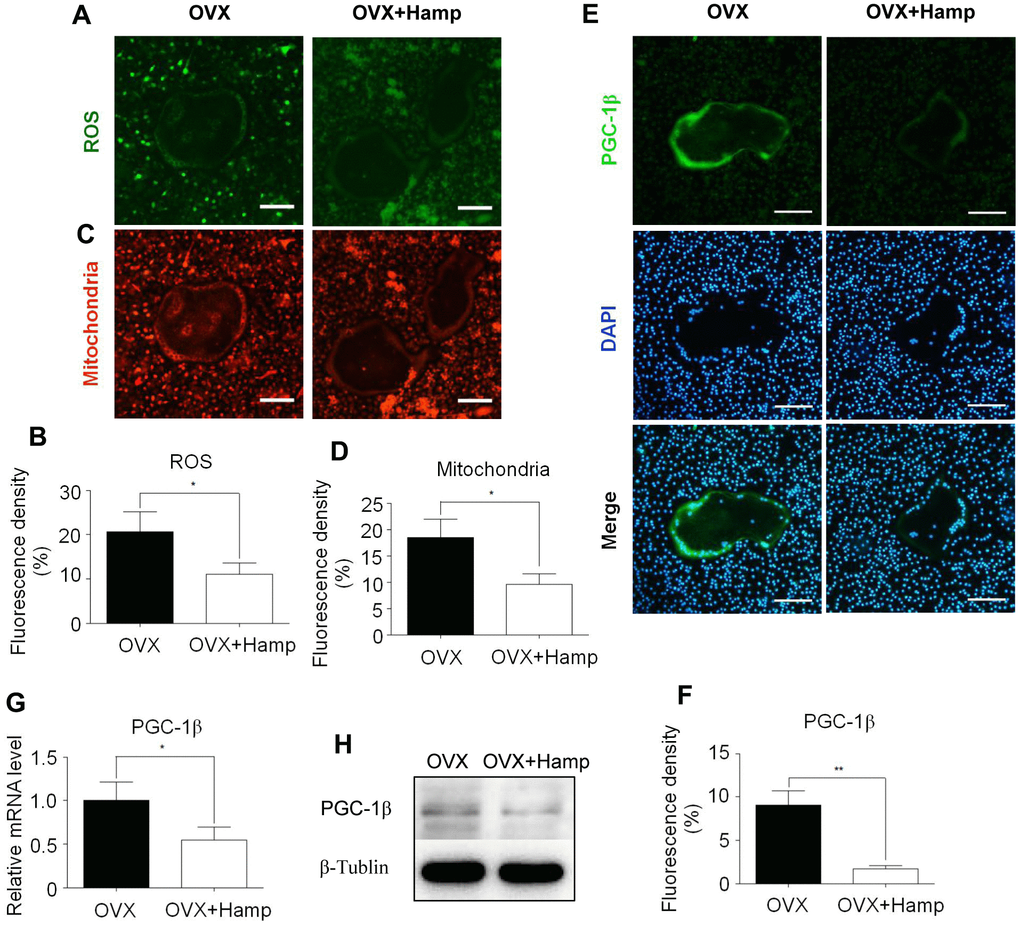
Figure 5.Hepcidin overexpression inhibits ROS production, mitochondrial biogenesis, and PGC-1β expression in osteoclasts. Bone marrow macrophages were extracted from femur and cultured with M-CSF and RANKL for 8 days. (A, C) The smaller cells are undifferentiated bone marrow macrophages and the larger cells in the middle are completely differentiated mature osteoclasts; (A) DCFH-DA and (C) mitochondrion-selective probes were used for assessing ROS and mitochondrial number in un-or-differentiated osteoclasts; (B, D) Mean fluorescence density of intermediate mature osteoclasts was measured to represent ROS and mitochondrial number respectively; (E) The smaller cells are undifferentiated bone marrow macrophages and the larger cells in the middle are completely differentiated mature osteoclasts; Immunocytofluorescence was used for assessing PGC-1β (E) in the osteoclasts, which were extracted from femur and cultured with M-CSF and RANKL for 8 days; (F) Mean fluorescence density of intermediate mature osteoclasts was measured to represent PGC-1β protein level; (G) the PGC-1β expression level was evaluated using qRT-PCR in osteoclasts; (H) PGC-1β protein levels were analyzed by western blotting in osteoclasts. Scale bar, 50 μm. The asterisks (*, **) indicate significant differences at P < 0.05, 0.01.