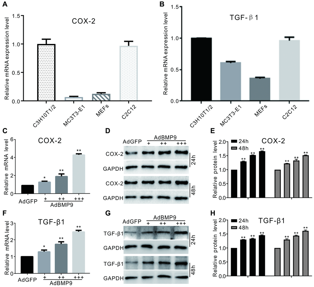
Figure 1.The effect of BMP9 on TGF-β1 and/or COX-2 in MSCs. (A) and (B) show real-time PCR analysis results of COX-2 or TGF-β1 expression in several kinds of progenitor cells. (C) COX-2 mRNA expression induced by BMP9 (real-time PCR). (D) Western blotting of the BMP9-induced protein level of COX-2. (E) Quantification of western blots showing the effect of BMP9 on COX-2 protein levels. (F) Real-time PCR data show that TGF-β1 was induced by BMP9. (G) Western blotting results show that TGF-β1 was induced by BMP9. (H) Quantification of western blotting shows that TGF-β1 was induced by BMP9. All experiments were performed in C3H10T1/2 cells. “+”, “++” and “+++” indicate increasing titer of recombinant adenovirus; “*”p < 0.05 and “**”p < 0.01.