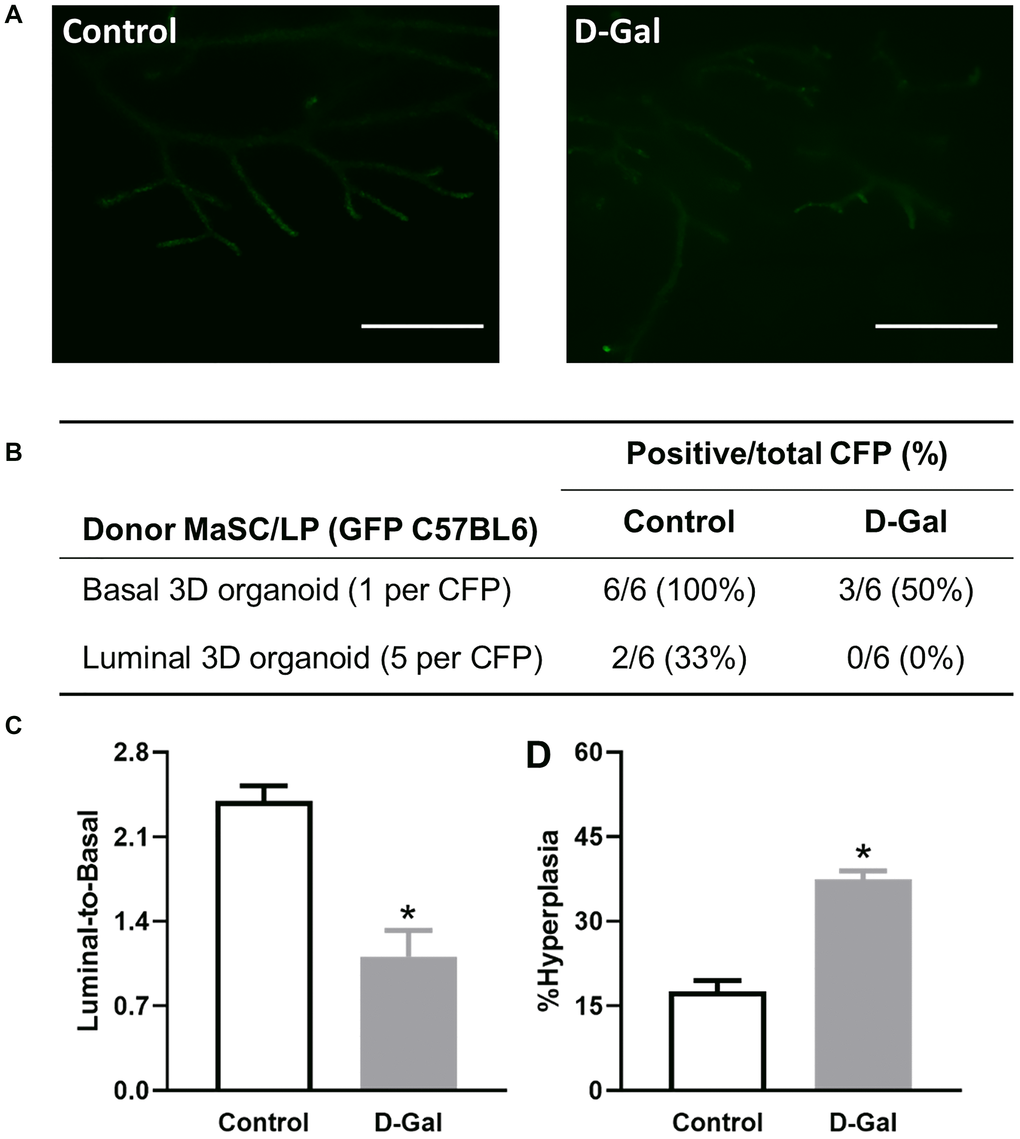
Figure 6.D-galactose alters mammary stem/progenitor cell function in vivo and increases hyperplasia in regenerated glands. (A) Regenerated glands from in vivo transplant of one basal 3D organoid from GFP mice with or without D-galactose treatment (scale bars, 1 mm); (B) Positive take of outgrowth from control or D-galactose-treated stem/progenitor cells in the cleared fat pad (CFP) transplant assay; (C) Luminal-to-basal cell ratio in mammary epithelial cells isolated from regenerated glands derived from control and D-galactose-treated GFP mice (n = 3–4); and (D) H&E histology analysis shows % hyperplasia in mammary ducts from regenerated glands derived from control and D-galactose-treated GFP mice (n = 3–6). Asterisks, significant difference between control and D-galactose (*P < .05).