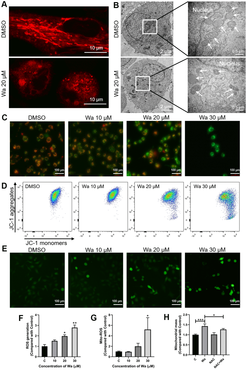
Figure 2.Warangalone causes to mitochondrial damage via ROS. (A, B) MDA-MB-231 cells were treated with 20 μM warangalone for 12 h. The mitochondria were stained with MitoTracker™ Red CMXRos 30 min, and observed by (A) confocal laser scanning microscopy (CLSM) (mitochondria emit red) and (B) transmission electron microscopy (TEM) (arrows mark mitochondria). (C, D) MDA-MB-231 cells were treated with the indicated concentrations of warangalone for 12 h. Fluorescence microscopy (C) and flow cytometry (D) were used to detect the mitochondrial membrane potential. JC-1 aggregates emit orange, and JC-1 monomers emit green. (e-g) MDA-MB-231 cells were treated with the indicated concentrations of warangalone for 12 h. Whole cell ROS was detected through fluorescence microscope (E) and fluorescence spectrophotometry (F). ROS labeled with DCFH-DA emit green (E). MitoSOX was used to specifically detect mitochondrial superoxides through flow cytometry (G). (H) MDA-MB-231 cells were pretreated with NAC (5 mM) for 1 h, and cultured with warangalone (20 μM) for 12 h. The mitochondria were stained with MitoTrackerTM Red CMXRos for 30 min and flow cytometry was used to detect mitochondrial mass. One-way ANOVA was used for statistical analysis (n ≥ 3). *P < 0.05, **P < 0.01, ***P < 0.001 compared to the respective control group.