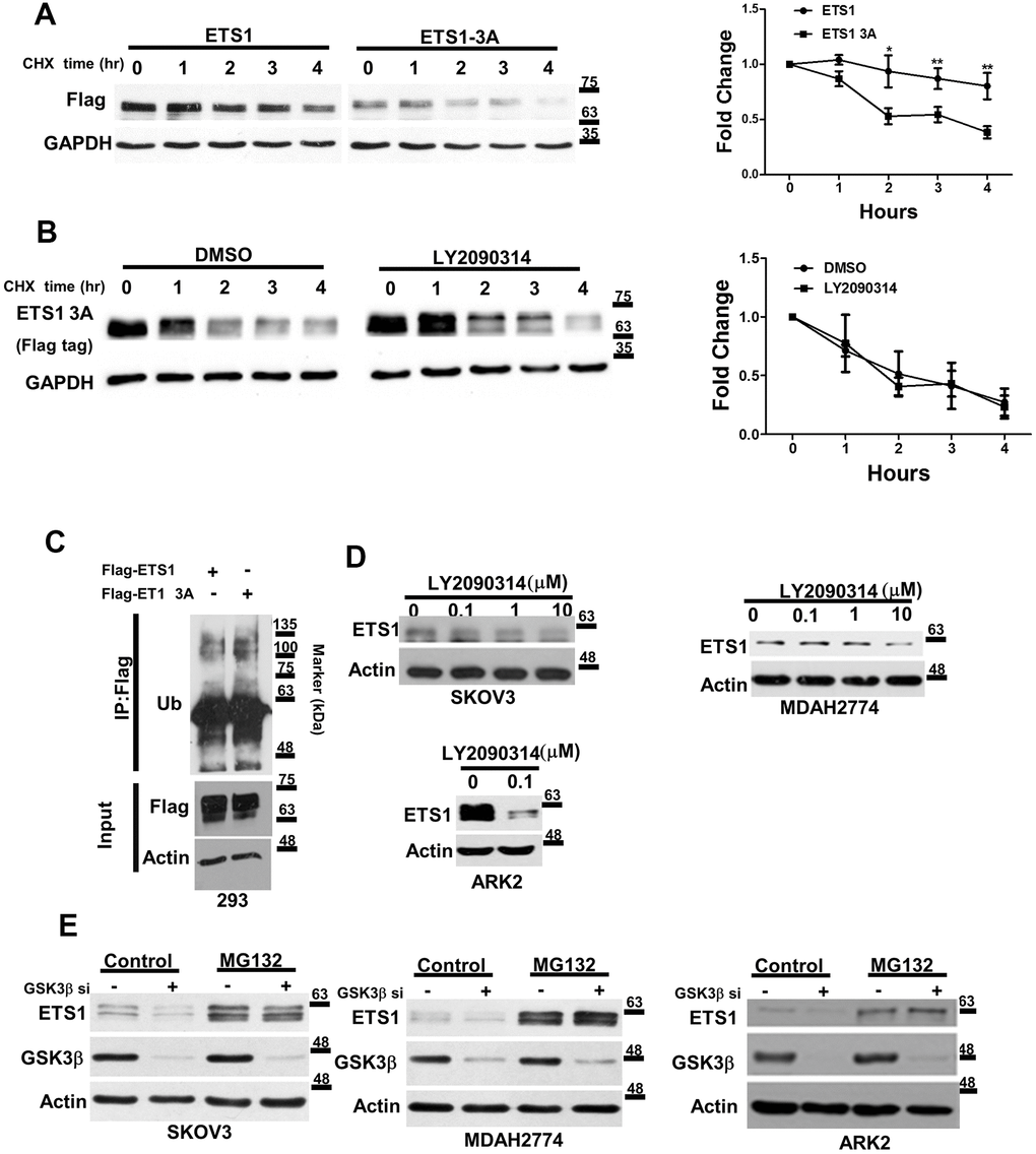
Figure 3.GSK3β regulates the stability of ETS1. (A) Flag-ETS1 and Flag-ETS1-3A were overexpressed in 293 cells and harvested at the reported time points in presence of cycloheximide (CHX). Flag-ETS1 and Flag-ETS1-3A protein levels were analyzed using an anti-Flag antibody. Endogenous GAPDH was used as a loading control, followed by normalization with an anti-GAPDH antibody. Fold changes in Flag-ETS1 or Flag-ETS1-3A protein levels are plotted in the right panel. Results are means ± standard errors of the mean from three independent experiments. *p < 0.05. (B) Flag-ETS1-3A were overexpressed in 293 cells and pre-treated with vehicle control or LY2090314 (0.1μM) for 1 h, finally harvested at the reported time points in the presence of cycloheximide (CHX). (C) Flag-ETS1 and Flag-ETS1-3A were overexpressed in 293 cells and harvested after treatment with MG132 (10 μM) for 5 h. Flag-ETS1 and Flag-ETS1-3A were precipitated with an anti-Flag antibody, whereas ubiquitin-bound proteins were detected with an anti-ubiquitin antibody. An Anti-Flag served as an input control and specific antibodies were used to detect Flag-ETS1, Flag-ETS1-3A, and actin. (D) The specific GSK3β inhibitor LY2090314 promoted ETS1 protein degradation. Ovarian cancer cells (SKOV3 and MDAH2774) and endometrial cancer cells (ARK2) were starved in OPTI-MEM medium in presence of the reported LY2090314 concentration for 24 h. Endogenous ETS1 and actin were analyzed with western blot using specific antibodies. (E) Ovarian and endometrial cancer cells were transfected with GSK3β siRNA for 72 h, and subsequently treated with MG132 (10 μM) or a vehicle for 5 h. ETS1, GSK3β, and actin protein levels were examined with western blot.