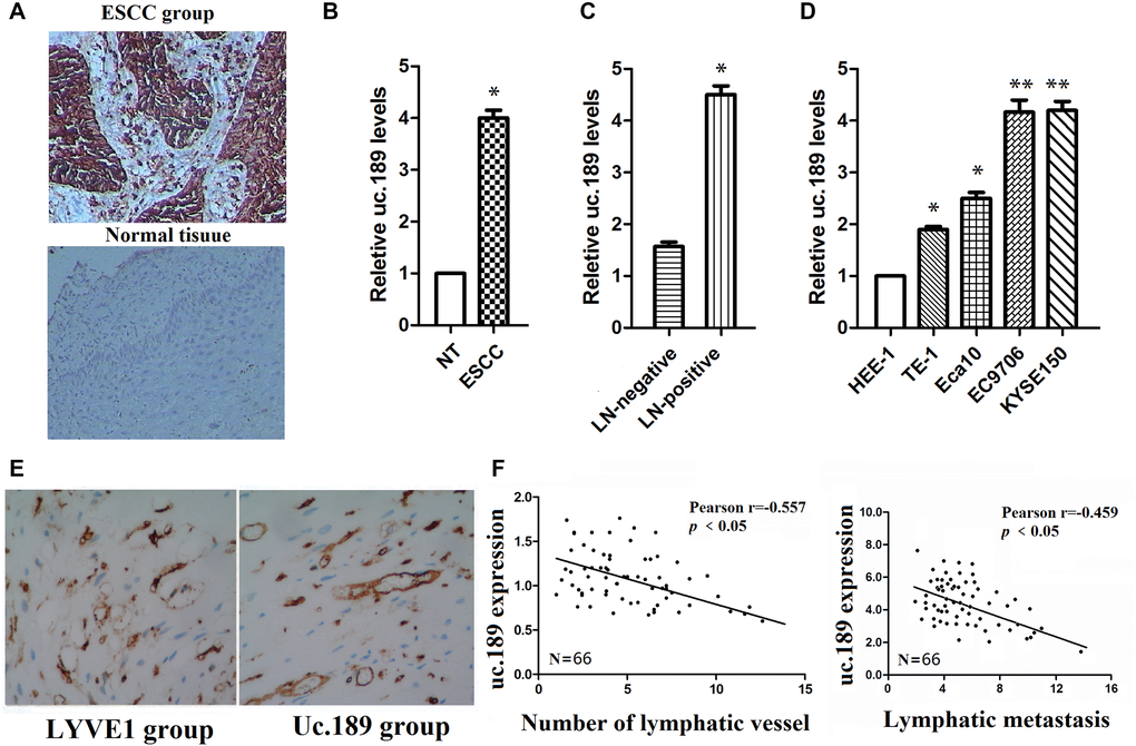
Figure 1.Uc.189 expressions are detected in ESCC tissues and cell lines. (A) uc.189 was a positive expression in ESCC and was a negative expression in normal tissue by ISH. (B) The uc.189 relative expression levels were determined by qRT-PCR in 66 paired human ESCC and normal tissue (NT) samples and normalized against an endogenous U6 RNA control. (C) The relative expression levels of uc.189 in ESCC with lymph-node metastasis (LN-positive; n = 58) and non-metastasis (NM; n = 8). (D) The relative expression levels of uc.189 in the four EC cell lines and normal cells (HEE-1). (E) uc.189 is a positive expression in the peritumoral lymphatic vessel (black arrow) of ESCC specimens and LYVE1 expression as a positive control. (F) Correlation analysis of ISH staining showing a negative correlation between uc.189 expressions and lymphatic vessel density indicated by anti-LYVE-S stain and uc.189 expressions are also a negative correlation with lymphatic metastasis in ESCC (n = 66). *p < 0.05, **p < 0.01.