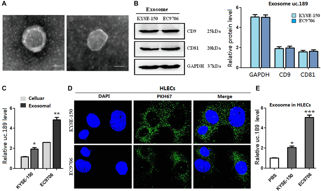
Figure 2.Exosomes of uc.189 can be secreted from ESCC cells and transferred to HLECs. (A) Photo of exosomes secreted from KYSE-150 and EC9706 was confirmed by transmission electron microscopy (Scale bar, 50nm). (B) Positive markers (CD9 and CD81) of KYSE-150 and EC9706-secreted exosomes were detected by western blot. (C) Uc.189 levels in ESCC cells and paired exosomes were detected by qRT-PCR. (D) HLECs pre-treated with PKH67-labeled exosomes for 48 hours were stained with DAPI (blue) for confocal microscopy analysis. Scale bar, 20 μm. (E) Uc.189 relative levels in HLECs pre-treated exosomes for 24 hours were detected by qRT-PCR. Error bars represent the mean ± SD of three independent experiments. *P < 0.05, **P < 0.01, ***P < 0.001.