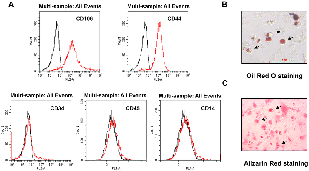
Figure 1.Characterization of DMSCs. (A) DMSCs were analyzed by FACS after staining with FITC- or PE-conjugated control isotype IgG (black peaks) or antibodies against the indicated cell-surface proteins. (B) DMSC differentiation. DMSCs were cultured in appropriate differentiation media to promote differentiation into adipocytes, as indicated by oil red O staining, and (C) osteoblasts, as indicated by alizarin red staining.