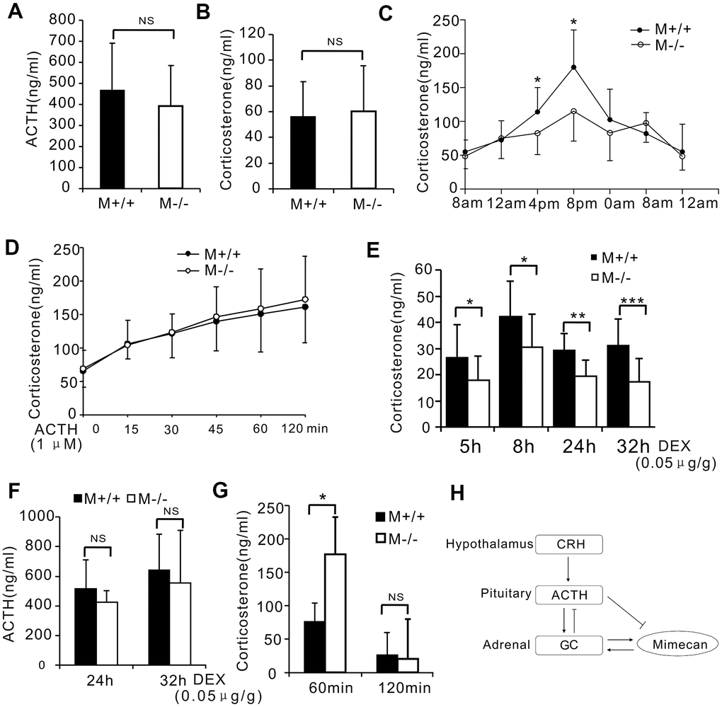
Figure 6.Disturbed stress-free diurnal rhythm of corticosterone secretion and hyperactivated stress response in mimecan-deficient mice. (A, B) No differences in serum corticosterone or plasma ACTH levels of wild-type (WT) and mim-/- mice were determined using ELISA (10 mice per group). (C) Significant disturbance of diurnal corticosterone secretion was observed in non-stressed mim-/- mice. Secretion levels lacked the typical peaks and troughs seen with WT mice (n=20 per group). (D) There was no difference in the serum corticosterone levels of wild-type (WT) and mim-/- knockout mice after stimulation with 1 μM ACTH. (E) The response to the DEX suppression test in mim-/- mice surpassed that of WT mice. The serum corticosterone level was determined using ELISA for 5–32 h after intramuscular injection of 0.05 ug/g DEX (21 mim-/- mice and 15 WT mice). (F) Similar serum ACTH levels in mim-/- and WT mice were observed 24–32 h after intramuscular injection of DEX (21 mim-/- mice and 15 WT mice). (G) A marked increase in the serum corticosterone level of mim-/- vs WT male mice was observed after 1 h of restraint and this was lost 1 h after release (5 mice per group). Data information: *p<0.05 for mim-/- vs. WT mice, Student’s t-test. (H) A graph showing the role of mimecan in the hypothalamic–pituitary–adrenal (HPA) axis.