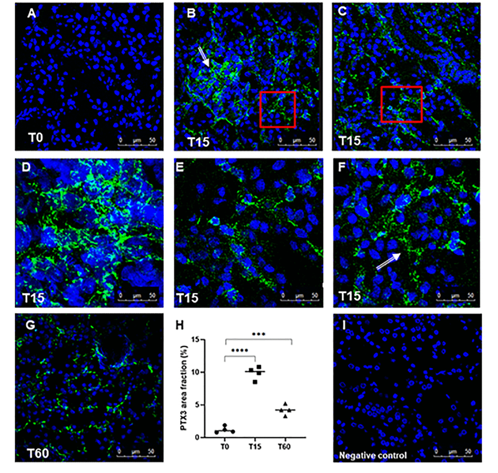
Figure 1.Analysis of PTX3 deposits in a swine model of I/R injury. Indirect immunofluorescence for PTX3 was performed on frozen pig kidney sections. A limited presence of PTX3 was observed in the biopsies at T0 (A). PTX3 deposits were observed after 15 min of reperfusion (B, C) at interstitial (E, zoomed image), peri-tubular (F, zoomed image) and glomerular (D) capillary levels. After 60 min the PTX3 deposits were still described at the level of peritubular capillaries (G). (I) Negative staining control for immunofluorescence was performed on cryosections with irrelevant primary antibodies for experimental conditions. Nuclei were highlighted with TO-PRO 3 in blue. Magnification 630X. (H) Quantification of PTX3 demonstrated a statistically significant increase after 15 min of reperfusion compared to basal biopsies. Results were expressed as % ± s.d. of positive area /high power field (hpf). *p<0.05 versus T0.