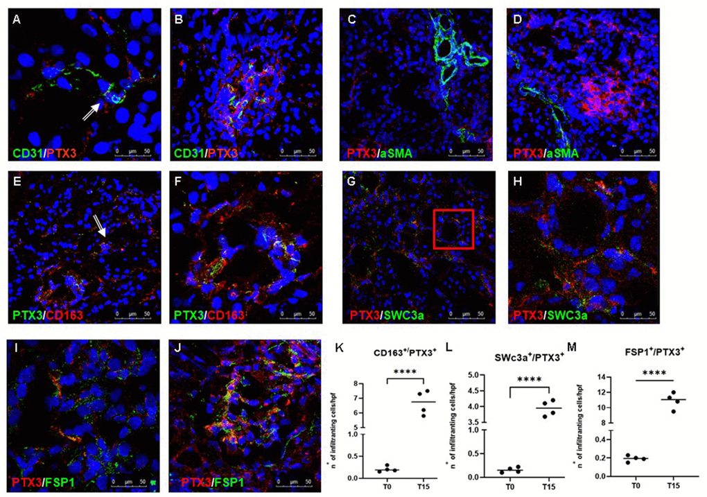
Figure 2.Characterization of the PTX3-associated cellular pattern in I/R injury. Frozen pig kidney sections were analyzed by indirect immunofluorescence to characterize the PTX3 source after 15 min of reperfusion. Co-localization between CD31 and PTX3 on renal EC was evident (A, B yellow staining). Activated myofibroblasts identified by alpha-smooth muscle actin (green) were negative for PTX3 (red; α-SMA+/PTX3-, C, D). Monocytes/macrophages identified by CD163 (red) co-localized with PTX3 (green; CD163+/PTX3+ yellow, (E) particular of E, F). Dendritic cells identified by SWC3a (green) were intensively positive for PTX3 (red; SWC3a+/PTX3+ yellow, (G) particular of G, H). Myofibroblasts identified by fibroblast-specific protein 1 (FSP1, red) co-localized with PTX3 (green; FSP1+/PTX3+ yellow, I, J). Nuclei were highlighted with TO-PRO 3 in blue. Original magnifications were x630. Quantification of CD163+/PTX3+ (K), SWC3a+/PTX3+ (L) and FSP1+/PTX3+ (M) cells demonstrated a statistically significant increase after 15 min of reperfusion compared to basal biopsies. Results were expressed as mean ± s.d. of infiltrating cells/high power field (hpf). *p<0.05 versus T0.