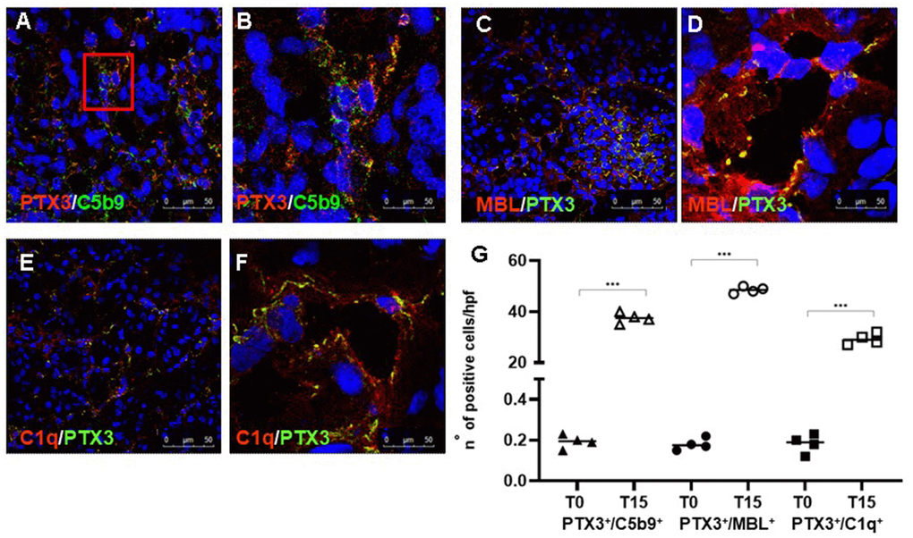
Figure 3.PTX3-mediated Complement activation in a pig model of I/R injury. Frozen pig kidney sections were examined by indirect immunofluorescence to investigate the co-localization (yellow staining) of C5b9 (green) and PTX3 (red) deposits (A, B). The co-localization between PTX3 (green) with MBL (red, C, D) and C1q (E, F) was investigated by immunofluorescence/confocal microscopy. PTX3 co-localized with MBL (C, D, yellow staining) and C1q (E, F, merge) at peri-glomerular (E) and peri-tubular (D, F) capillary sites. In confocal microscopy images nuclei were stained with TO-PRO 3 (blue). (G) Quantification of C5b9+/PTX3+, MBL+/PTX3+and C1q+/PTX3+cells compared to basal biopsies. Results were expressed as % ± s.d. of positive area /high power field (hpf). *p<0.05 versus T0.