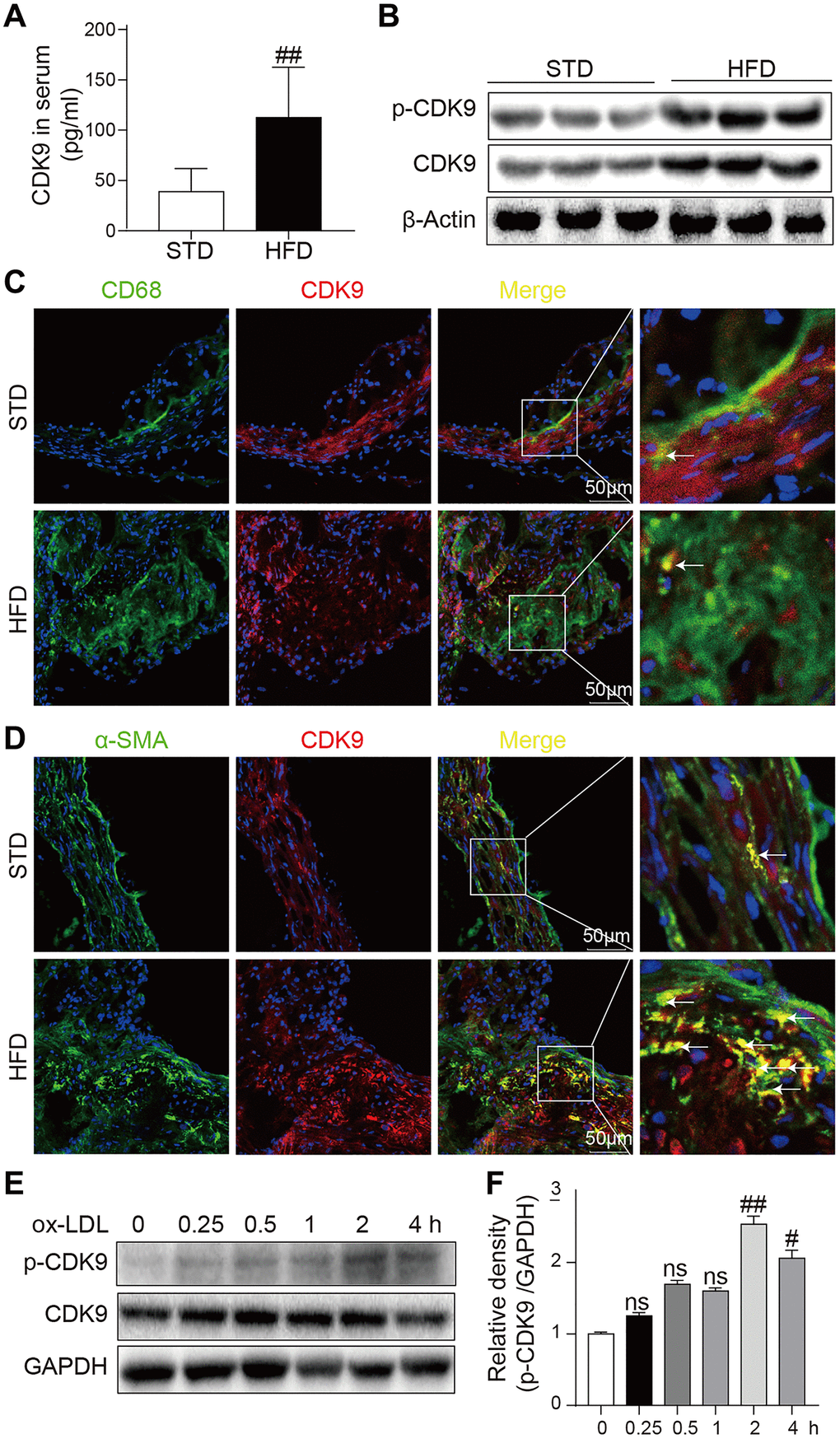
Figure 1.CDK9 is expressed in VSMCs in atherosclerotic lesions. (A) Serum levels of CDK9 were detected using ELISA assay (n = 8; **P < 0.01 compared to STD). (B) p-CDK9 and CDK9 protein levels in aortas of ApoE-/- mice were detected by western blotting. (C) Representative immunofluorescence staining of CDK9 (red) and macrophage marker CD68 (green). Tissues were counterstained with DAPI (blue). Yellow arrows indicate co-location of CDK9 and CD68 stanning (scale bar = 50 μm). (D) Representative immunofluorescence staining of CDK9 (red) and VSMCs marker α-SMA (green). Tissues were counterstained with DAPI (blue). Yellow arrows indicate co-location of CDK9 and α-SMA stanning (scale bar = 50 μm). (E, F) Western blot analysis of p-CDK9 and CDK9 protein levels in VSMCs challenged with 50 μg/ml ox-LDL for the indicated time points (n = 8; ##P < 0.01 compared to 0h).