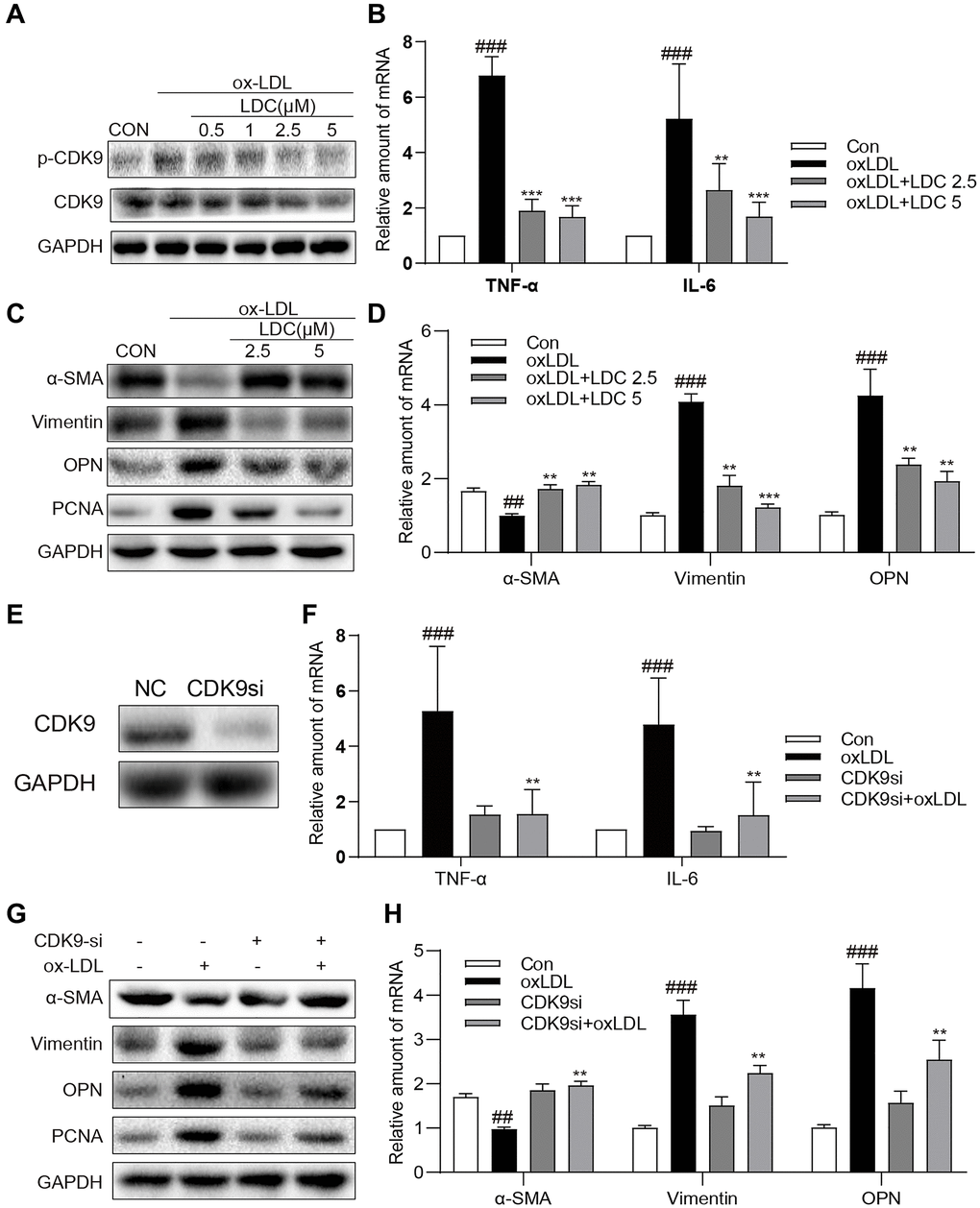
Figure 4.Inactivation of p-CDK9 by CDK9 inhibitor or CDK9-si prevented ox-LDL-induced inflammation and phenotype switch of VSMCs in vitro. (A) LDC000067 suppressed ox-LDL-induced activation of p-CDK9. VSMCs were pretreated with LDC000067 (indicated concentrations) for 1 h and then incubated with ox-LDL (50 μg/mL) for 2 h. The levels of p-CDK9 and CDK9 were detected by western blot. (B–D) VSMCs were treated with LDC000067 (2.5 or 5 μM) for 1 hour and then exposed to ox-LDL (50 μg/mL) for 6 h (in panels B), 24 h (in panels C) or 12 h ((in panels D). (B) The levels of TNF-α and IL-6 were detected using real-time qPCR assay. (C) Expressions of α-SMA, Vimentin, OPN and PCNA in the cultural medium were detected by western blot. (D) The mRNA levels of α-SMA, Vimentin, OPN were detected using real-time qPCR assay (n = 3 ##P < 0.01, ###P < 0.001, compared to control; *P < 0.05, **P < 0.01, ***P < 0.001, compared to ox-LDL). (E–H) VSMCs were transfected with siRNA against CDK9 and then incubated with ox-LDL (50 μg/mL) for 6 h (in panels G), 24 h (in panels H) or 12 h ((in panels H). (E) VSMCs were transfected with siRNA against CDK9 for 6 h and then detected expression of CDK9 by western blot. (F) The level of TNF-α and IL-6 were detected using real-time qPCR assay. (G) Expressions of α-SMA, Vimentin, OPN and PCNA in the cultural medium were detected by western blot. (H) The mRNA level of α-SMA, Vimentin, OPN were detected using real-time qPCR assay (n = 3; ##P < 0.01, ###P < 0.001 compared to control; *P < 0.05, **P < 0.01, compared to ox-LDL).