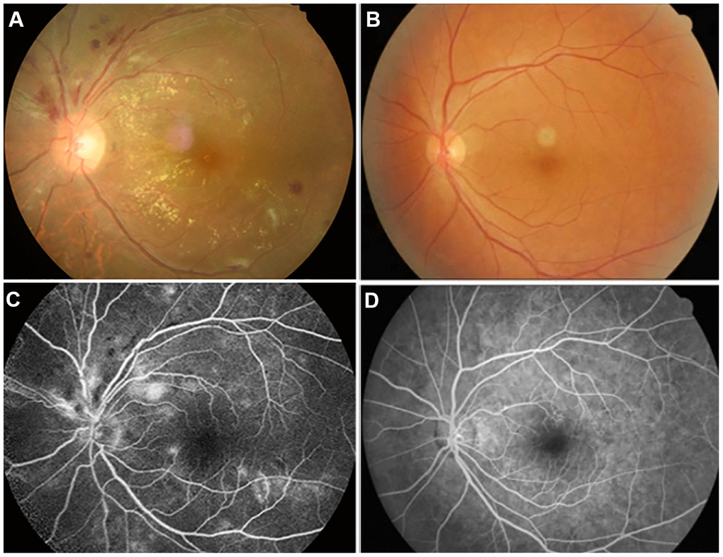
Figure 1.Examples of retinal fundus photography (above) and fluorescence fundus angiography (below) in the HR patients and HC group. Notes: (A) shows the left retinal fundus photos of patients with hypertensive retinopathy and (B) shows the left retinal fundus photos of normal people. (C) shows left fluorescence fundus angiography in patients with hypertensive retinopathy, and (D) shows corresponding fluorescence fundus angiography in normal subjects. (A, C) Correspond to the same person, and (B, D) correspond to another person.