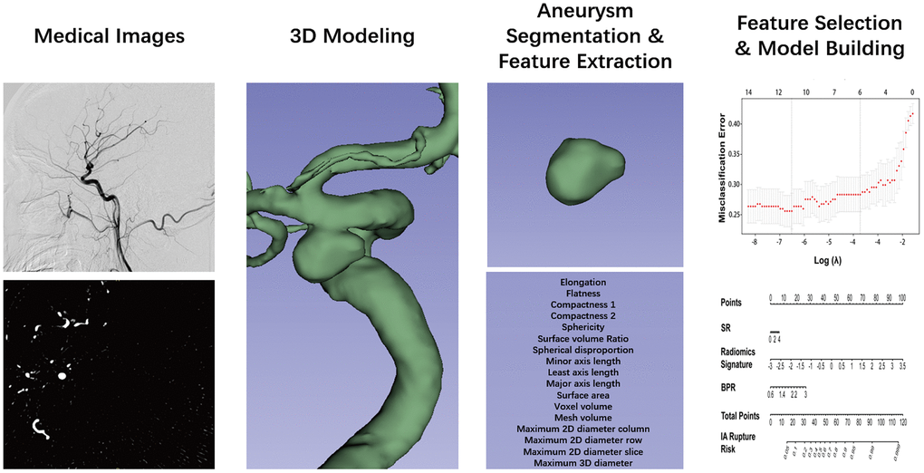
Figure 2.Flow chart of the study. The aneurysm was reconstructed from DSA images and using 3D slicer. The segmentation was performed by threshold and checked layer by layer. Then, the segmented label map and volume files were entered in the Pyradiomics package in the Python platform, and 17 radiomics morphological features were extracted for each aneurysm. The least absolute shrinkage and selection operator binary logistic were used to select the potential assessment factors and develop a radiomics signature. Along the radiomics morphological features, 16 traditional morphological features were combined and entered in the model construction analysis. Finally, the optimal model was performed in the nomogram.