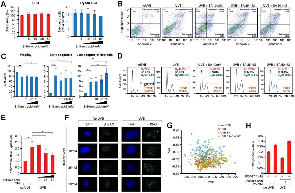
Figure 4.(A) SRB and Trypan blue cell viability assays performed in cells treated with or without Shikimic acid (5, 10, 25 and 50 mM) for 24h. (B) Apoptosis detection using the Annexin V-FITC/PI double staining followed by flow cytometry in non-irradiated and non-treated cells, irradiated and non-treated cells and irradiated cells treated with Shikimic acid (10, 25 and 50 mM). (C) Representation and statistical analysis of the percentage of apoptotic cells in each of the conditions analyzed. (D) Cell cycle analysis using PI staining followed by flow cytometry. (E) Relative p16INK4a mRNA levels monitored by qPCR in the indicated conditions. Student T-test, *p<0.05. (F) Immunofluorescence of γH2AX in non-irradiated and non-treated cells, irradiated and non-treated cells and irradiated cells treated with Shikimic acid (10, 25 and 50 mM). (G) Principal component analysis (PCA) on Savitzky–Golay second derivatized spectra in the fingerprint region (1800–950 cm−1) for non-irradiated cells (No UVB), irradiated non-treated cells (UVB), irradiated cells treated with Shikimic acid 25 mM (UVB SA) and irradiated cells treated with Shikimic acid 25 mM plus EX-527 1 μM (UVB SA+EX-527). Student T-test, *p<0.05, **p<0.01, ***p<0.001. (H) Beta-sheet to alpha-helix ratio obtained by curve-fitting analysis of the amide I band (1700-1600 cm-1).