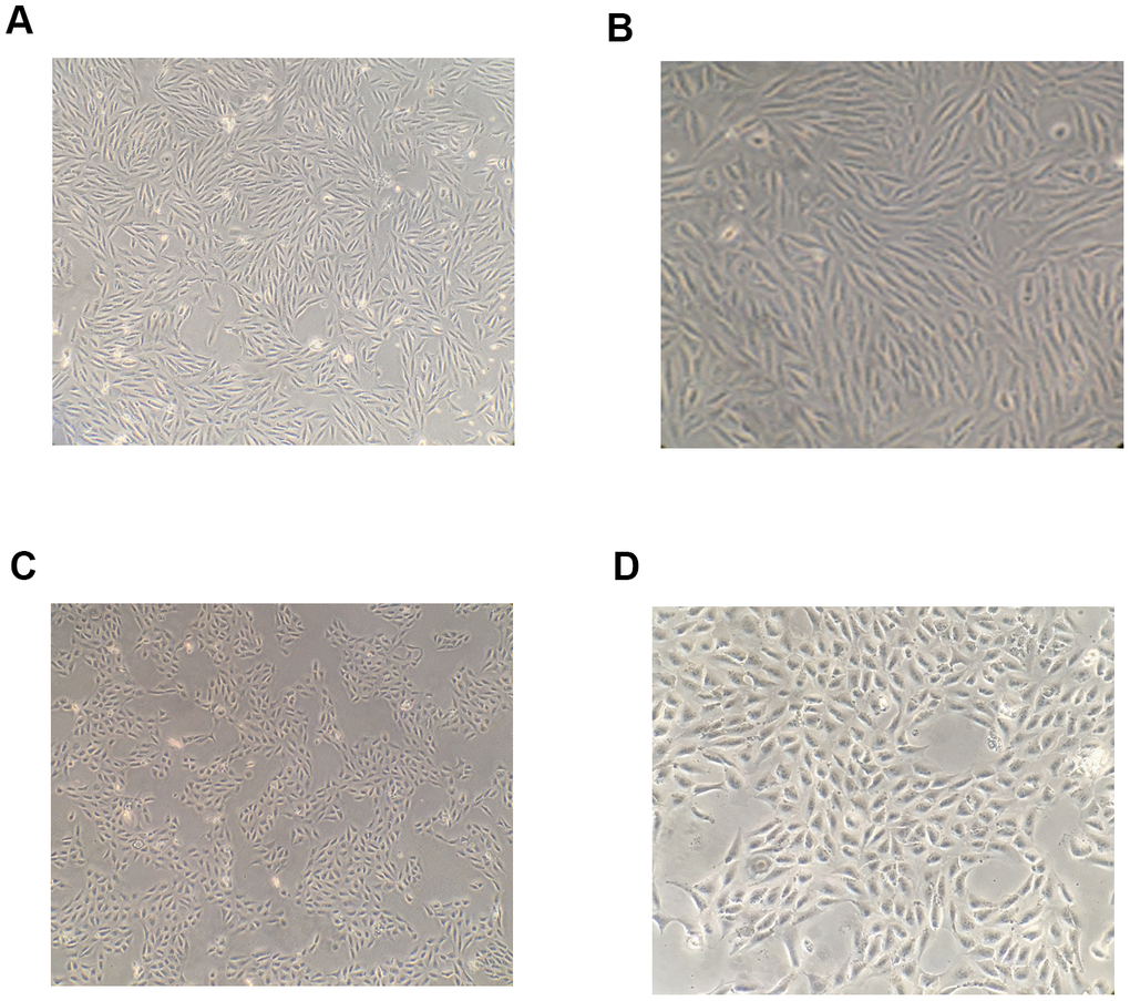
Figure 1.Morphological changes of RPE cells following induction by TGF-β1. After 48-h induction with 10 ng/mL TGF-β1, the shape of RPE cells became spindle-like and their arrangement was observed to become loose under a microscope (A, B) The shape of RPE cells in the blank control group under a microscope (C, D).