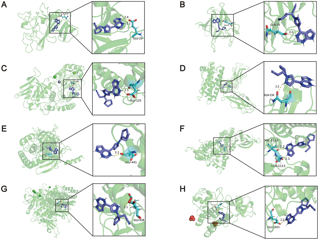
Figure 6.The results of the molecular docking simulations. (A) There were two hydrogen bonding between the amino acid residue of ANGPT2 (PDB: 4JZC) (GLU29) and ruxolitinib, and the distance between the atoms was 2.1Å and 2.4Å. (B) The amino acid residue of FGF7 (PDB: 1QQL) bound to ruxolitinib was GLN35, and the distance was 2.0Å. (C) The amino acid residue of NT5E (PDB: 4H2F) bound to ruxolitinib was GLU125, and the distance was 2.1Å. (D) The amino acid residue of CSF3R (PDB: 2D9Q) bound to ruxolitinib was GLN234, and the distance was 2.2Å. (E) The amino acid residue of JAK1 (PDB: 4GS0) bound to ruxolitinib was GLU441, and the distance was 2.1Å. (F) The amino acid residues of JAK2 (PDB: 2B7A) bound to ruxolitinib were VAL1110 and ASN1111, and the distance were 2.2Å and 2.1Å. (G) The amino acid residue of JAK3 (PDB: 3ZC6) bound to ruxolitinib was GLU938, and the distance was 2.1Å. (H) The amino acid residue of TYK2 (PDB: 4GFO) bound to ruxolitinib was GLU1053, and the distance was 2.2Å.