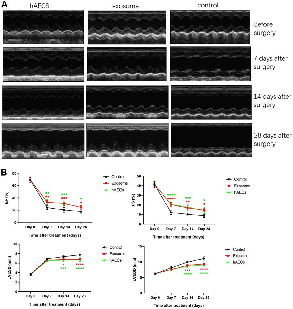
Figure 4.Echocardiography shows that cardiac function is improved by implanting hAECs and their exosomes in a rat acute myocardial infarction model. (A) Representative M-mode echocardiography of SD rats before and after MI in the hAEC treatment group, exosome group and control group. (B) The line charts shows the analysis of the left ventricular ejection fraction (EF), left ventricular fractional shortening (FS), left ventricular end-diastolic diameter (LVEDD) and left ventricular end-systolic diameter (LVESD) in SD rats 7, 14 and 28 days after MI, respectively. The data are expressed as the means ± SD, n=8 per group, *P<0.05.