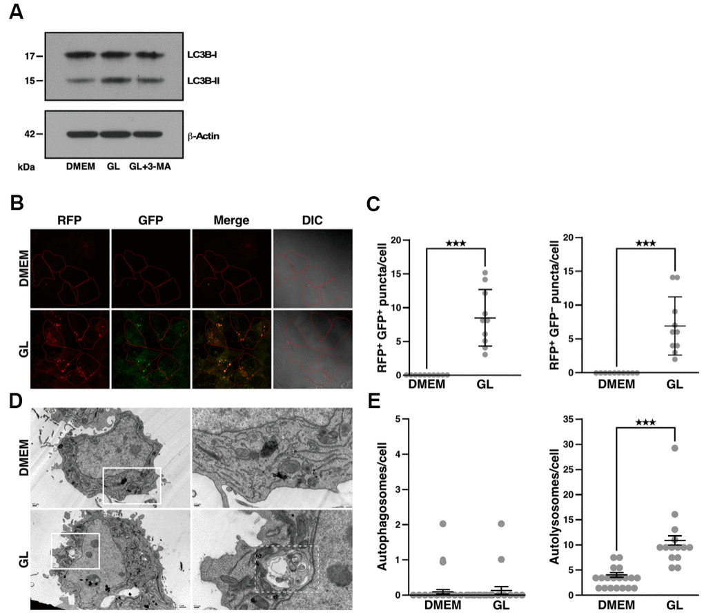
Figure 4.G. lucidum induces autophagy in human cells. (A) Effects of G. lucidum (GL) on LC3B-I and LC3B-II in human cells. Huh7 cells maintained in Dulbecco’s modified Eagle’s medium (DMEM) were treated with GL (1%) or DMEM for 12 hrs, prior to treatment with DMEM containing 3-methyladenine (3-MA, 2 mM) for 12 hrs. Protein levels were monitored by Western blot and normalized against actin. (B) GL induces the formation of autophagosomes and autolysosomes in human cells. Huh7 cells expressing monomeric red fluorescent protein (RFP)-LC3 and green fluorescent protein (GFP)-LC3 were treated with GL (1%) for 24 hrs, prior to fluorescence microscopy analysis. In differential interference contrast (DIC) images, cells are delineated in red for clarity. (C) Quantification of fluorescent puncta based on fluorescence microscopy. (D) Representative transmission electron microscopy (TEM) images of GL-treated cells. Huh7 cells were treated with GL (1%) for 24 hrs prior to fixation and thin-sectioning as described in Materials and Methods. Images on the right correspond to the insets delineated by white rectangles in the images on the left. An autolysosome is delineated by a white dashed line for the GL panel. (E) Quantification of autophagosomes and autolysosomes based on TEM. ***p<0.001.