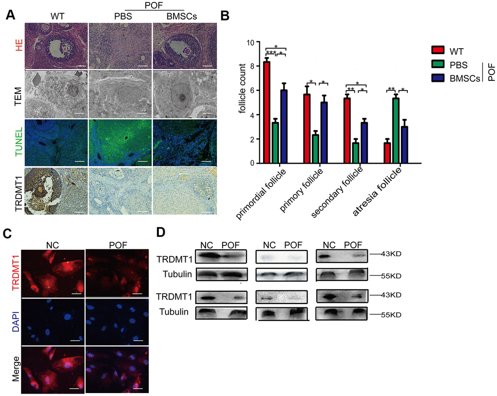
Figure 1.Reduced TRDMT1 is associated with decreased ovarian function. (A) Histopathological examination of the ovaries of the WT, POF-PBS and POF-BMSCs groups. Scale bar: 100 μm. (B) Primordial follicles, primary follicles, secondary follicles, and atretic follicles were observed. (A) Transmission electron microscopy analysis of the ovarian structures in each group of mice. Scale bar: 2μm. (A) Apoptosis of GCs in ovaries was measured by TUNEL staining. Representative images of terminal deoxynucleotidyl transferase dUTP nick end labeling (TUNEL) staining show apoptotic GCs in each group. The TUNEL-positive apoptotic cells are indicated by green fluorescence. The nuclei (blue) were stained with DAPI. Scale bar: 100μm. (A) Representative immunohistochemical images of TRDMT1 in each group. Brown staining represents a positive TRDMT1 signal. Scale bar: 100μm. (C) Representative immunofluorescent staining of TRDMT1 in GCs from women with normal ovarian function and from patients with POF. Nuclei were stained with DAPI. Scale bar: 50μm. (D) TRDMT1 expression in the serum of healthy people and patients with POF was detected by Western blotting. An anti-Tubulin antibody was used as a loading control. *p<0.05, **P<0.01, and ***P<0.001. Statistical significance was determined using two-tailed t-tests for two groups and ANOVA for multiple comparisons. All values are means ± SD.