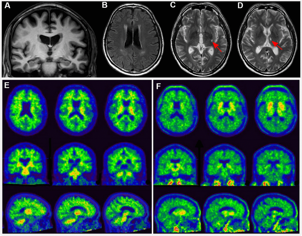
Figure 1.Clinical utility of AD-RAI in MCI subjects. A 68-year-old man with 11 years of education had complaints of memory decline for over 3 years. Z-score in Trial 4 of HKLLT was -1.94 SD (≤ -1 SD, i.e. MCI). Visual MTA rating score on MRI was 1 (≥ 1), (A) which was suggestive of AD. However, HV measures yielded conflicting results, with HF of 0.47% (> 0.41%) and raw HV of 7.38ml (> 6.07mL) suggestive of non-AD. FLAIR and T2-weighted sequences showed periventricular white matter hyperintensity and two subcortical lacunes (red arrows) (B–D). AD-RAI was only 0.11 (< 0.5) also suggestive of non-AD. Subsequent PIB PET (E) and T807 PET (F) showed negative results (i.e. A-T-), supporting the finding of AD-RAI. The MCI syndrome and mild MTA might be associated with cerebral SVD (i.e. vascular MCI associated with SVD).