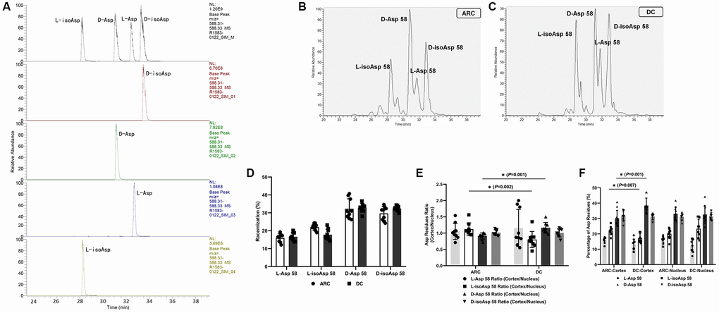
Figure 1.(A) Representative LC-MS/MS trace showing the separation of the four Asp isomers of the αA-crystallin tryptic peptide (55–65) TVLDSGISEVR. Peptides containing D-Asp, D-isoAsp, L-Asp, or L-isoAsp at position 58 were synthesized. To measure racemization in αA-crystallin, all forms of the peptide were summed and modifications for each were expressed as a% of the total peak area. (B) Representative graphs showing the separation of the four Asp 58 isomers in αA-crystallin of ARC lenses. (C) Representative graphs showing the separation of the four Asp 58 isomers in αA-crystallin of DC lenses. (D) The percentage of each Asp 58 isomer in αA-crystallin from lenses of patients with ARC and DC. (E) The cortex/nucleus ratio of each Asp 58 isomer in αA-crystallin from cortex and nucleus of ARC and DC lenses after dissection. (F) The percentage of each Asp 58 isomer in αA-crystallin from cortex and nucleus of ARC and DC lenses after dissection.