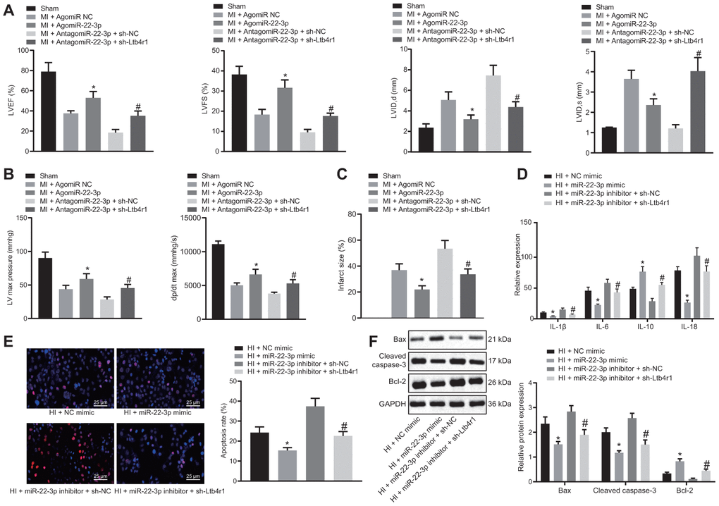
Figure 3.miR-22-3p contributes to alleviation of CHD-related myocardial injury by downregulating Ltb4r1. MI mice injected with AgomiR-22-3p or AntagomiR-22-3p and sh-Ltb4r1. (A) Quantification of LVEF, LVIDD, LVIDs, LVEF, LVFS, in myocardial tissues of MI mice. (B) Hemodynamic analysis of LV and dP/dt in myocardial tissues of MI mice. (C) The infarct size in myocardial tissues of MI mice detected using TTC staining upon treatment with MI cardiomyocytes treated with exogenous miR-22-3p mimic or miR-22-3p inhibitor and sh-Ltb4r1. (D), Levels of IL-1β, IL-6, and IL-18 in hypoxia-induced MI cardiomyocytes measured using ELISA. (E), Representative images of apoptosis of hypoxia-induced MI cardiomyocytes detected by TUNEL staining (× 400). (F) Protein levels of Cleaved caspase-3, Bax, and Bcl-2 in hypoxia-induced MI cardiomyocytes determined using Western blot analysis, normalized to GAPDH. * p < 0.05 vs. MI + NC mimic and # p < 0.05 vs. AntagomiR-22-3p + sh-NC or MI + miR-22-3p inhibitor + sh-NC. Data among group were analyzed by one-way ANOVA/Tukey’s test.