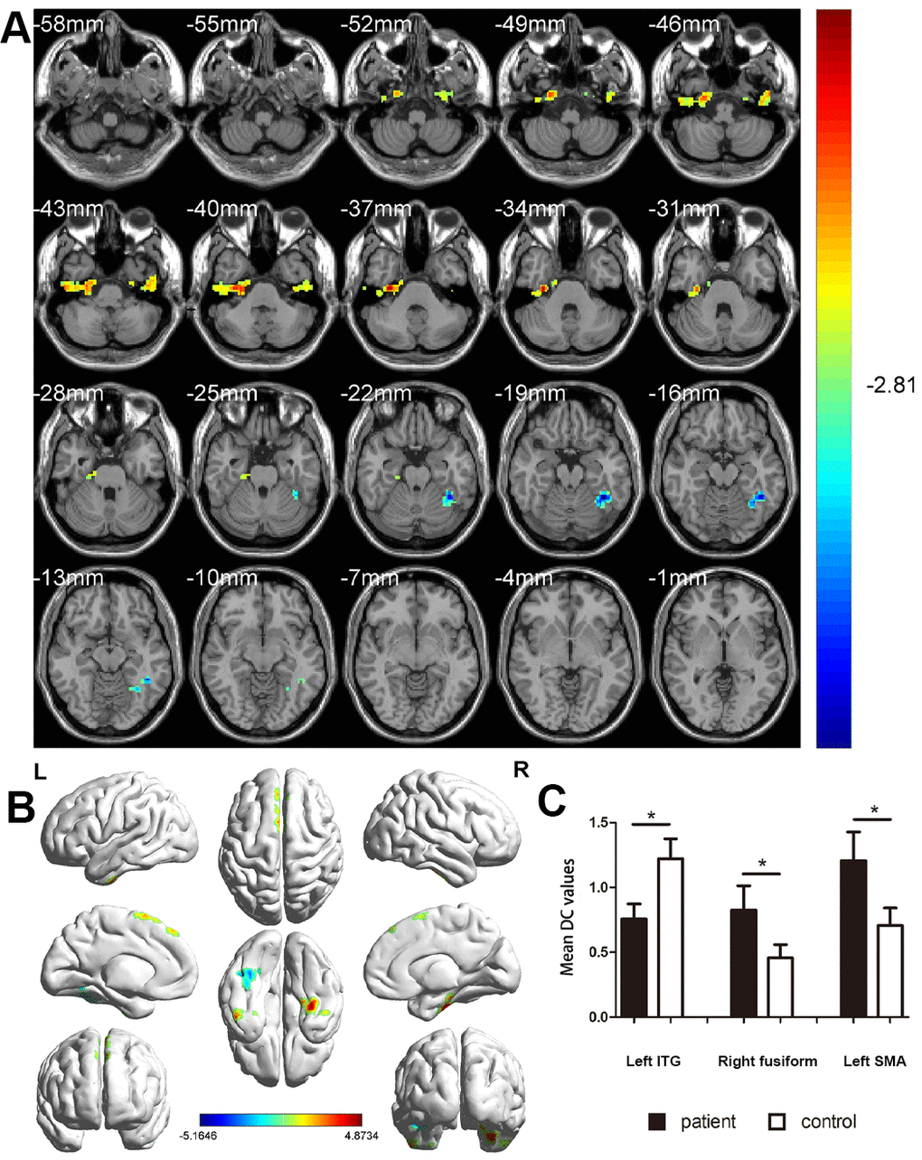
Figure 2.Comparison of DC values in MCI and HC groups. (A) Differences in DC were found in left ITG, right fusiform gyrus, and left SMA. (B) The stereoscopic form of the cerebrum. The red area indicates an increase in DC value; the blue indicates a decrease in DC value. (GRF correction, the cluster-level: P<0.05; two-tailed, with voxel level P<0.005). (C) The Mean DC value between MCIs and control group. Abbreviations: DC, Degree centrality; MCI, mild cognitive impairment; HC, healthy controls; ITG, inferior temporal gyrus; SMA, supplementary motor area.