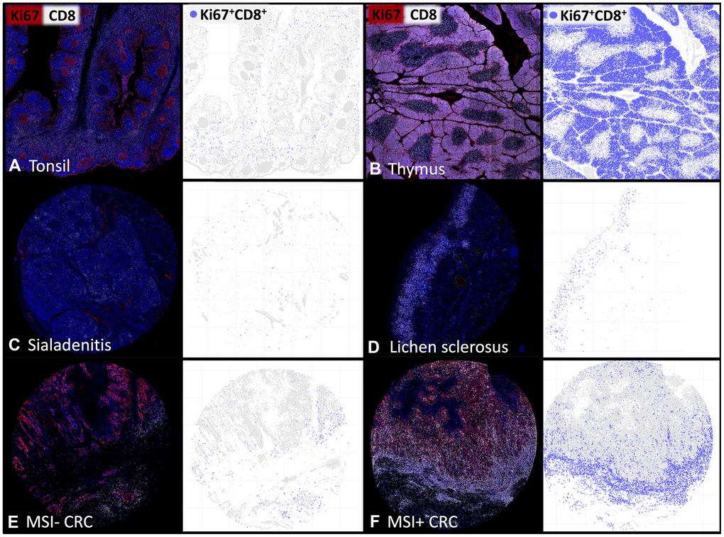
Figure 2.The density of proliferating CD8+ T-lymphocytes varies between tissues and individual patients. Representative multiplex immunofluorescence images of CD8+ (white) and Ki67+ (red) cells in (A) normal human tonsil, (B) thymus, (C) sialadenitis, (D) lichen sclerosus, (E) Microsatellite stable colorectal cancer (MSI- CRC) and (F) Microsatellite instable colorectal cancer (MSI+ CRC). The visualizations of the digital image analysis highlight the subset of Ki67+CD8+ proliferating cytotoxic T-cells (blue).