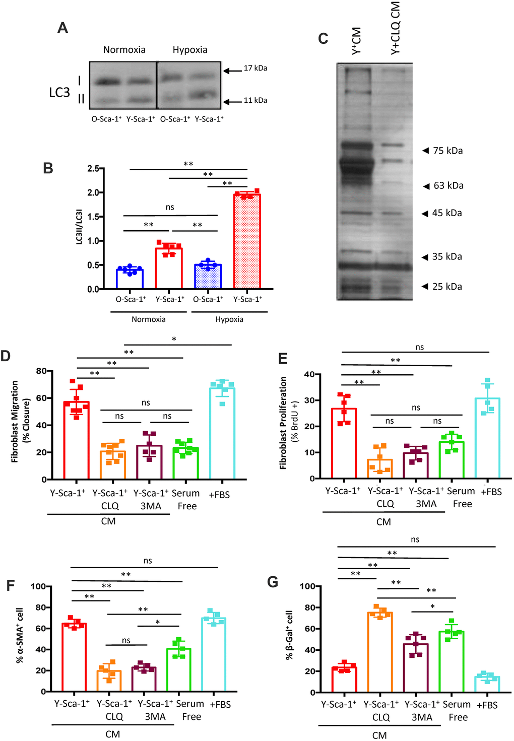
Figure 2.Autophagy in Y-Sca-1+ BMCs is associated with beneficial paracrine effects on old cardiac fibroblasts. (A) LC3II and LC3I were measured in Y-Sca-1+ and O-Sca-1+ bone marrow cells (BMCs) under normoxic (n=6) and hypoxic (n=4) conditions. (B) Quantification of LC3II/I protein intensities shown in panel (A). (C) Conditioned medium (CM) was harvested from Y-Sca-1+ (Y+CM) and Y-Sca-1++ chloroquine (CLQ, Y+CLQ CM). Proteins were separated by 10% SDS-PAGE. Representative image of silver-stained gel from Y+CM±CLQ. (D–G) CM was collected from Y-Sca-1+ BMCs treated with chemical inhibitors of autophagy, CLQ and 3-Methyladenine (3MA), and added to cultured cardiac fibroblasts from old mice (>20 months old) for 48 hours. Treatments are abbreviated as: Y-Sca-1+ CLQ and Y-Sca-1+ 3MA, serum free (serum-free media), +FBS (complete growth media). (D) Percent wound closure (after completing scratch wound assay) was measured using ImageJ. (E) Proliferation was assessed as the percentage of BrdU+ cells, normalized to total cell number. (F) Differentiation was determined as the percentage of α-SMA+ cells, normalized to total cell number. (G) Senescence was measured as the percentage of β-galactosidase+ cells, normalized to total cell number. Data with multiple groups were analyzed using one-way ANOVA, while data with two groups were analyzed by t-test. **p≤0.01; ns: not statistically significant.