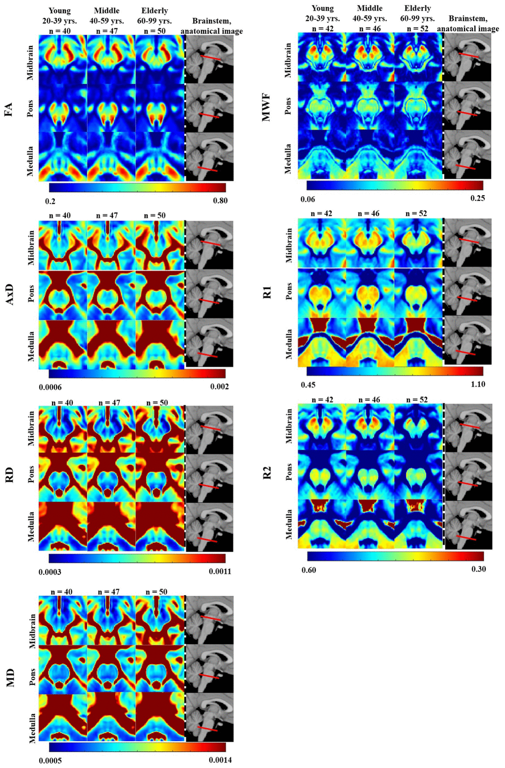
Figure 1.FA, AxD, RD, MD, MWF, R1, and R2 represented as averaged participant maps calculated for three age groups. Three representative slices covering respectively the midbrain, pons, and medulla are displayed. The red bars on the anatomical images indicate the location of these slices. Visual inspection indicates an increase in R1, R2, and MWF from early adulthood, 20-29 years, through middle age, followed by a decrease in several brainstem substructures, and a more generally monotonic decrease in FA. Inspection of AxD, RD, and MD demonstrated a slight decrease from early adulthood through middle age followed by an increase in several brainstem substructures.