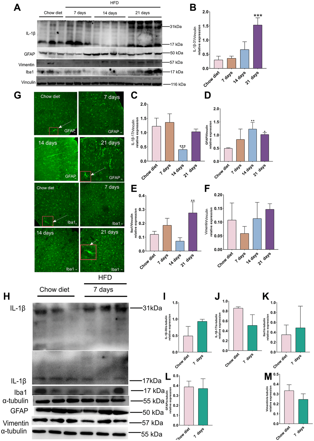
Figure 4.Effect of HFD on activation of astrocytes and microglia in the mice cortex and cerebellum. (A) Western blots and (B–F) densitometric analysis for IL-1β 31 kDa (B), IL-1β 17 kDa (C), GFAP (D), Iba-1 (E), vimentin (F) after HFD in mice cerebral cortex at day 7, day 14 and day 21 (n = 3 to 4 per group). (G) Immunohistochemistry of GFAP+ astrocytes and Iba-1+ microglia in cerebral cortex tissue sections. High magnification images of astrocytes and microglia in a larger box outlined by red color indicated by arrows. Magnification, ×40. Scale bars, 10 μm. (H) Western blot analysis of cerebellum tissues of mice with HFD and chow diet controls (n = 3 per group). (I–M) Quantification of IL-1β 31 kDa (I), IL-1β 17 kDa (J), Iba-1 (K), GFAP (L) and vimentin (M).Vinculin and β-actin as a loading control. Values are presented as means ± SD. *P < 0.05 and **P < 0.01 versus chow diet; one-way ANOVA by Tukey’s test for cerebral cortex. Two-tailed Student’s t test for cerebellum.