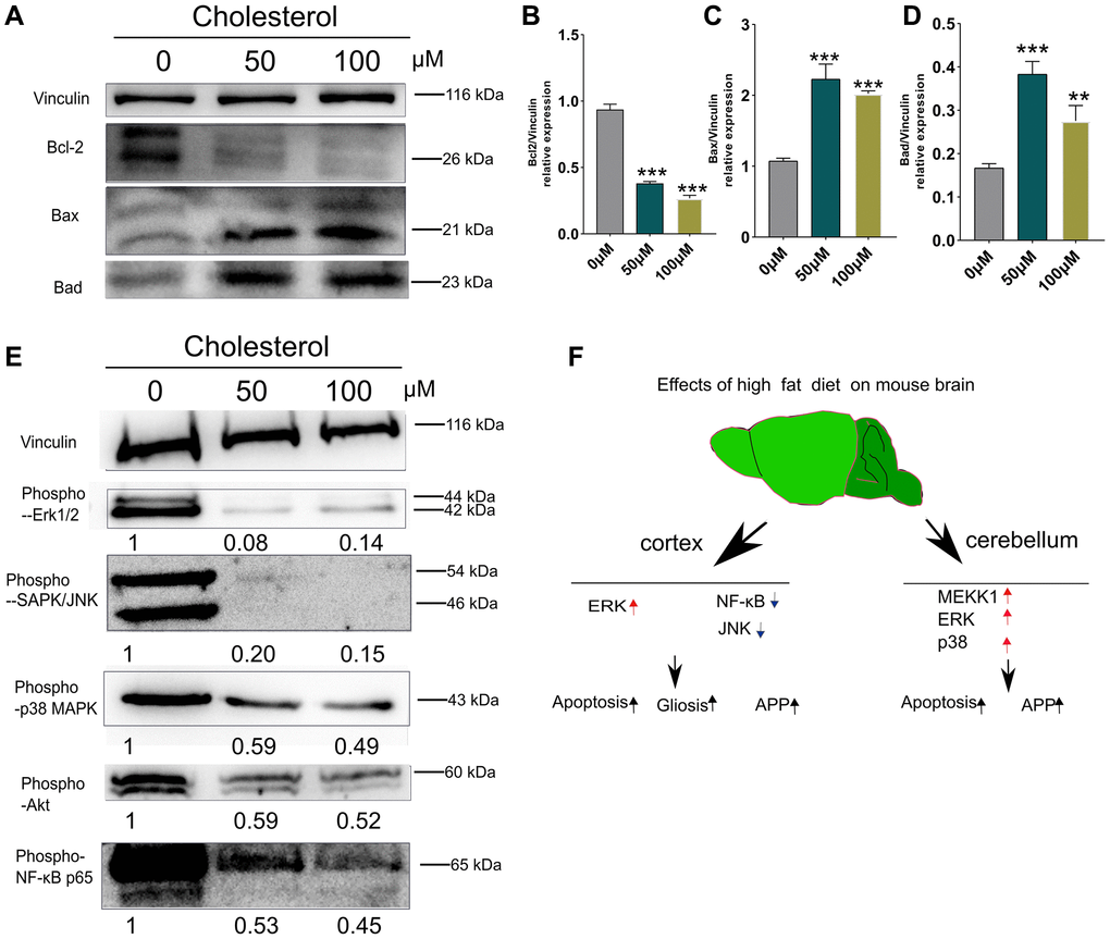
Figure 6.Changes of apoptosis and signaling pathways induced by cholesterol in SH-SY5Y cells. SH-SY5Y Cells were treated with cholesterol at indicated concentration (0 μM, 50 μM, and 100 μM) for 48 h (n = 3 per group). (A) Western blots and (B–D) Quantitation for Bcl-2 (B), Bax (C), and Bad (D) in the supernatants of SH-SY5Y Cells. (E) Western blots of phospho-p44/42 MAPK (Erk1/2), phospho-SAPK/JNK, phospho-p38 MAPK, phospho-Akt, phospho-NF-κB p65 from supernatants of SH-SY5Y Cells after 48 h culture. The levels of phosphorylated signaling-related proteins after treatment with 50 μM, and 100 μM cholesterol were normalized to the levels of the 0μM treatment group. (F) Diagram showing the mechanism of how short-term HFD aggravates apoptosis, glial cell activation and APP production in cerebral cortex and cerebellum. Vinculin as a loading control. Values are presented as means ± SD. *P < 0.05 and **P < 0.01 versus chow diet; one-way ANOVA by Tukey’s test. Red arrow indicates up-regulation and Black arrow indicates down-regulation.