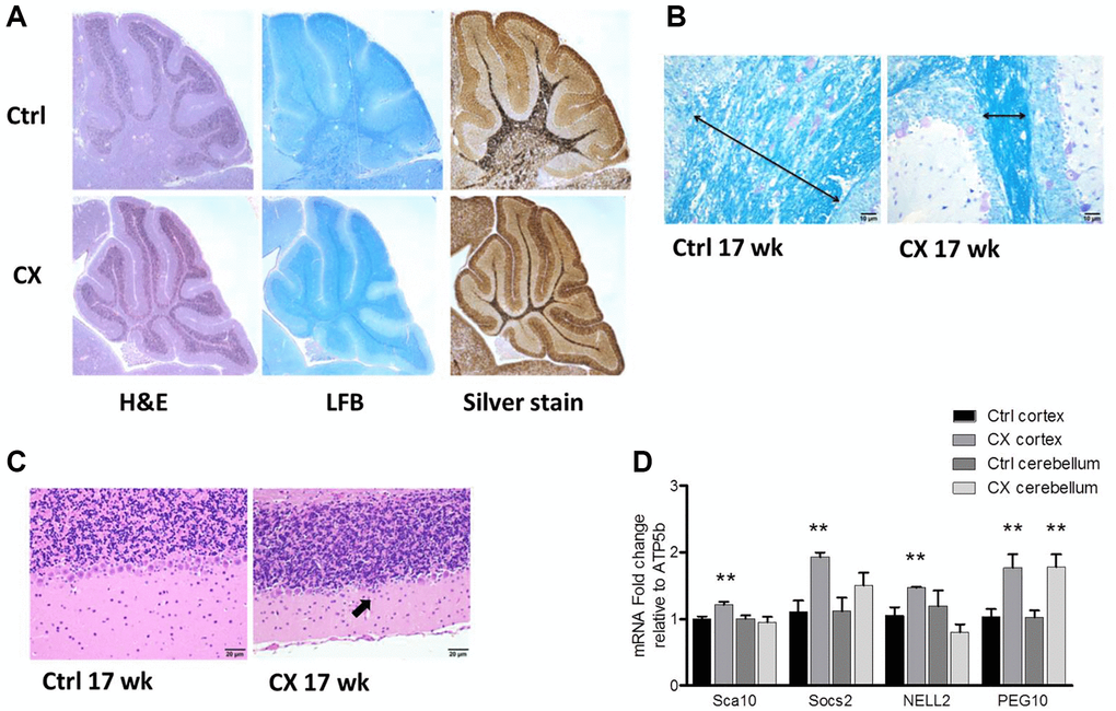
Figure 1.Neurodegeneration and myelin loss in CX mice. (A) Hematoxylin and Eosin (H&E), Luxol fast blue (LFB), and silver staining of cerebellum of pre-weaning control and CX animals. (B) Luxol fast blue staining of cerebellum from 17-week old control and CX animals. Myelin content length indicated by arrow. (C) Hematoxylin and Eosin staining of the Purkinje cell layer of animals described in b. CX mice display degeneration of the Purkinje cell layer (arrow). n = 2. (D) Gene expression of control and CX cortex and cerebellum for PARP-1 regulated genes, n = 8. Data are presented as mean ± SE. Student’s t-test. *P < 0.05, **P < 0.01. Abbreviations: Ctrl: control; WT: wildtype; Wk: weeks.