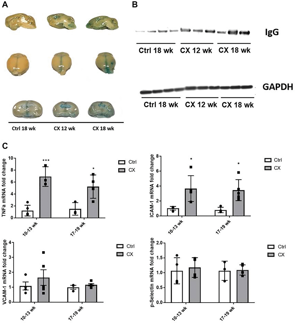
Figure 2.CX mice display brain vascular permeability and expression of vascular cell activation genes. (A) Evans blue stain shows higher permeability in CX brains in an age-dependent manner (n = 2). (B) Western blot of IgG, another marker of permeability of the BBB in mice brain. GAPDH was used as a loading control. (C) qRT-PCR of vascular cell activation markers, with ICAM-1 and TNFα in CX mice being significantly higher than control Csa−/− mice, in both 10–13 and 17–19 week old age groups, though there was no difference between age groups. Data are presented as mean ± SE. Two way Anova followed by Tukey’s post hoc test, n ≥ 4. *P < 0.05, **P < 0.01, ***P < 0.001 when comparing within the same age group. Abbreviations: Ctrl: control; Wk: weeks.