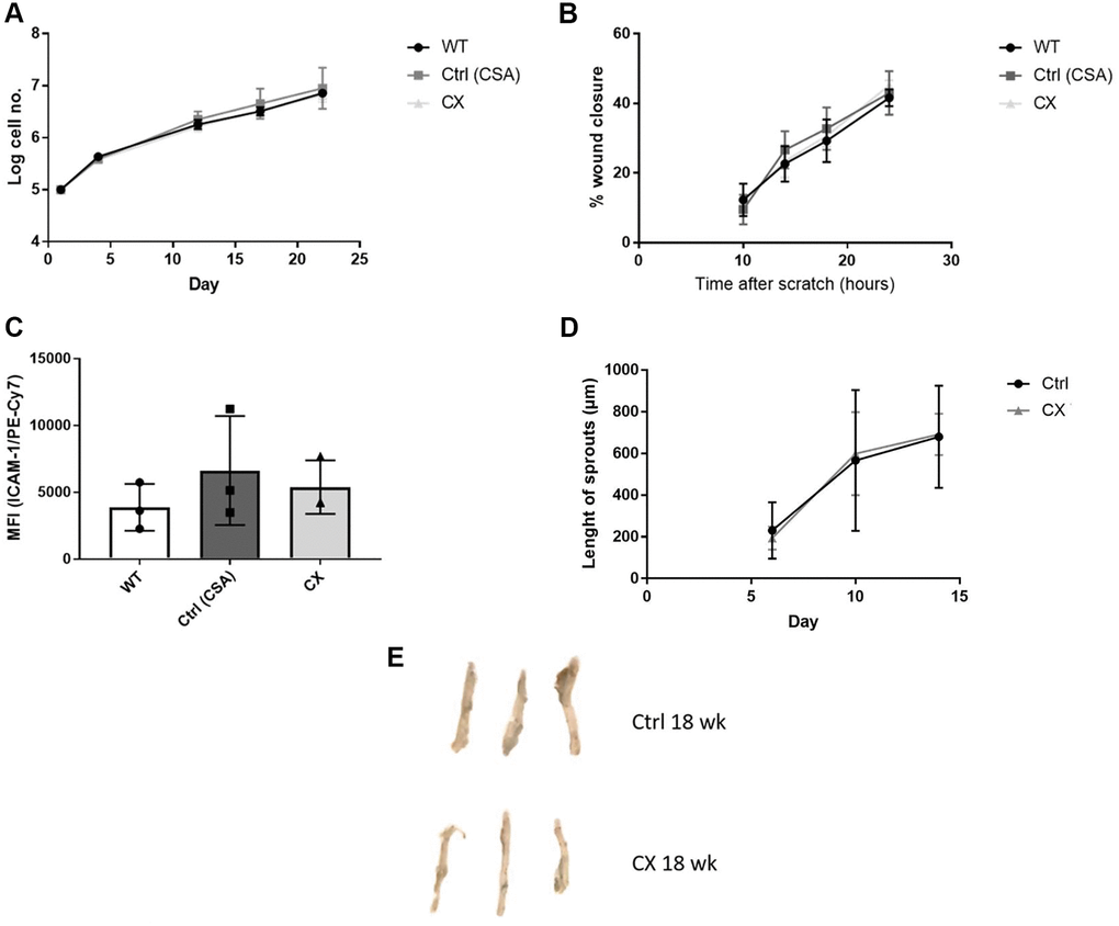
Figure 3.CX mice do not display cell autonomous vascular dysfunction phenotype regarding endothelial cell proliferation, migration, ICAM-1 activation, angiogenesis nor senescence. (A) Proliferation rates of CX ECs do not differ from neither the CSA KO nor WT ECs. (B) Migration capacity, as measured by the wound healing assay. (C) ICAM-1 expression in ECs, measured by fluorescence intensity in FACS analysis. One-way Anova followed by Tukey’s post hoc test. (D) Angiogenesis capacity, measured using the aortic ring assay in 12 week old animals. Data are presented as mean ± SE. Student’s t-test. p values of higher than 0.05 were considered nonsignificant. (E) Senescence Associated β-galactosidase staining of mice aortas. n ≥ 3 for all experiments. Abbreviations: Ctrl: control; WT: wildtype; EC: endothelial cells; Wk: weeks.