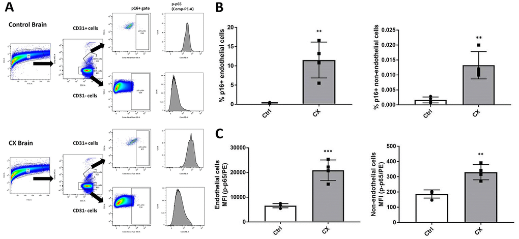
Figure 4.CX mice brain display p16Ink4a and p-p65 upregulation in endothelial and non-endothelial cells. (A) Flow cytometry gating strategy for the detection of senescent and pro-inflammatory endothelial and non-endothelial cells. Whole brain cells were stained for EC marker CD31, senescence marker p16Ink4a, and active NF-κB (phosphorylated p65). Brain cells were first identified using a forward scatter (FSC) and side scatter (SSC) gate. CD31 positive and negative cells were then identified by their APC fluorescence levels. p16+ senescent cells were then identified using an SSC and Alexa Fluor 488 gate. Phosphorylated-p65 expression in CD31+ and CD31- cells was measured by analyzing PE median fluorescence intensity. (B) FACS analysis of endothelial and non-endothelial p16Ink4a positive cells in CX mice brains. (C) Phosphorylated p65 FACS analysis of endothelial and non-endothelial cells in CX mice brains, n = 4. Data are presented as mean ± SE. Student’s t-test. *P < 0.05, **P < 0.01. Abbreviation: Ctrl: control.