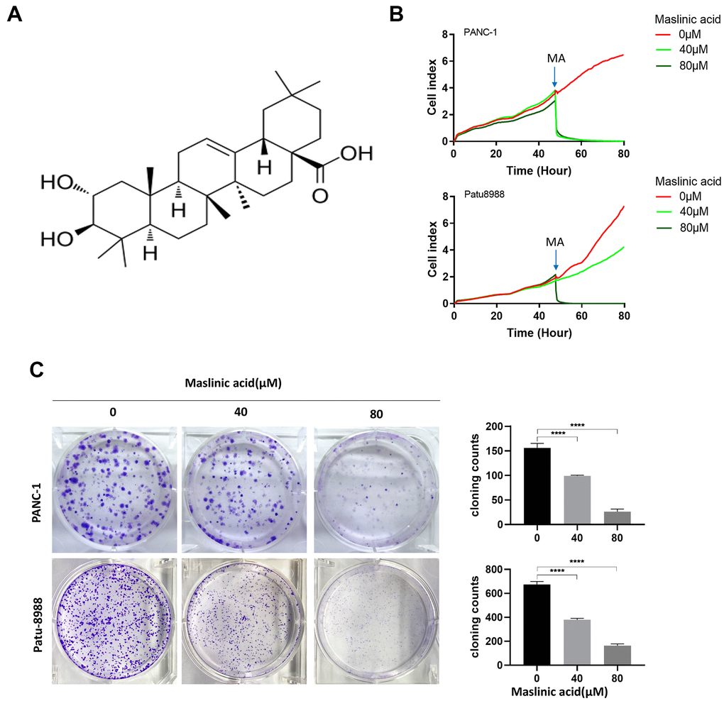
Figure 1.Maslinic acid (MA)-induced inhibition of the proliferation of PANC-1 and Patu-8988 cells. (A) The chemical structure of MA. (B) Label-free Real-time Cellular Analysis (RTCA) of PANC-1 and Patu-8988 cells incubated with MA (0, 40, 80 μM). (C) The effects of MA on the colony formation of PANC-1 and Patu-8988 cells. The cells were exposed to MA (0, 40, 80 μM) for 24 h. After 14 days cells were stained with crystal violet and colony counted. Data are representative of three independent experiments, expressed as mean ± SD. *p <0.05, **p < 0.01, ***p < 0.001, ****p < 0.0001.