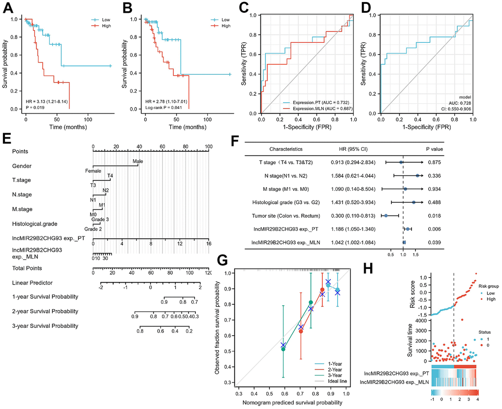
Figure 9.Prognostic model based on lncRNA MIR29B2CHG expression in CRC patients. Kaplan–Meier analysis of Disease-free survival(DFS) of CRC patients based on lncRNA MIR29B2CHG expression in primary tumor tissue (A) and in lymphnodal metastasis tumor tissue (B). Data are shown as hazard ratios (95% CI). (C) Time-dependent ROC curve for assessing the prognostic accuracy of CRC patients by the lncRNA MIR29B2CHG expression in primary tumor and lymphnodal metastasis tumor tissue, separately. (D) Time-dependent ROC curve for assessing the prognostic accuracy of CRC patients by the joint expression of lncRNA MIR29B2CHG in both primary tumor and lymphnodal metastasis tumor tissue. Data are shown as AUC (95% CI). ROC = receiver operator characteristic. AUC = area under the curve. (E) The nomogram was utilized by adding up of the points identified on the points scale for each variant. The total points occurred on the bottom scales represent the probability of 1-, 2- and 3-year survival. (F) Univariate analysis was performed in CRC cohort. The bar corresponds to 95% confidence intervals. (G) The calibration curve based on the expression of lncRNA MIR29B2CHG in primary tumor and lymphnodal metastasis tumor tissue for predicting DFS at 1-, 2- and 3-years in CRC cohort. (H) The distribution of risk score of the established prognostic model by lncRNA MIR29B2CHG expression.