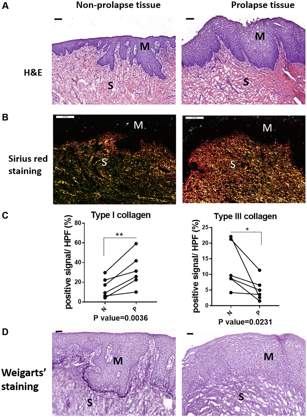
Figure 1.The micro-structural changes in vagina tissue of pelvic organ prolapsed patients. (A) HE staining revealed obvious microstructure porosity and disappeared Vicious Angle structure in the prolapsed tissue (M: mucous layer, S: submucosa layer. scale bar 100 um), (B) Sirius red staining showed increased type I collagen and decreased type III collagen in the prolapsed tissue (type I collagen was red and bright yellow, type III collagen was green, scale bar 200 um), (C) Sirius red staining as in B was quantified, the proportion of positive signals area (red signals for type I collage and green signals for type III collagen) in each High Power Field of vision (HPF) were quantified, n = 6 in each group. (D) Weigarts’ staining demonstrated disappeared elastic fibers near basal layer in the prolapsed tissue (scale bar 50 um).