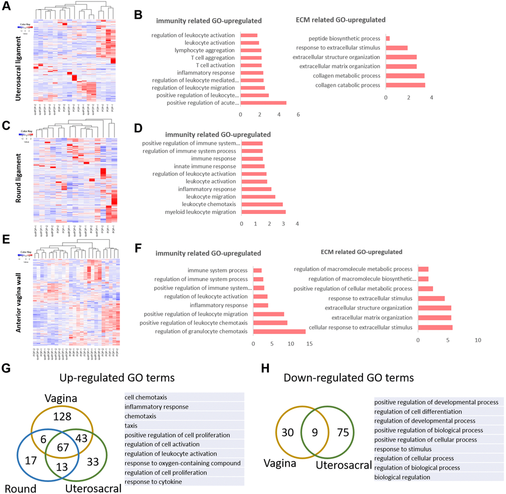
Figure 3.The molecular changes in pelvic tissue of pelvic organ prolapsed patients. (A, B) Heat map across all the samples using the top 500 most differently expressed genes (A) and gene ontology (GO) categories (B) of up-regulated differently expressed genes in uterosacral ligament between the prolapsed group and non-prolapsed group. (C, D) Heat map across all the samples using the top 500 most differently expressed genes (C) and gene ontology (GO) categories (D) of up-regulated differently expressed genes in round ligament between the prolapsed group and non-prolapsed group. (E, F) Heat map across all the samples using the top 500 most differently expressed genes (E) and gene ontology (GO) categories (F) of up-regulated differently expressed genes in vagina tissue between the prolapsed group and non-prolapsed group. (G) Venn diagram showing the overlaps of up-regulated differentially expressed genes enriched GO terms between vagina, round ligament and Uterosacral ligament, the top 10 GO terms also showed on the right side. (H) Venn diagram (left) and the form (right) showed the overlaps of down-regulated differentially expressed genes enriched GO terms between vagina and Uterosacral ligament.