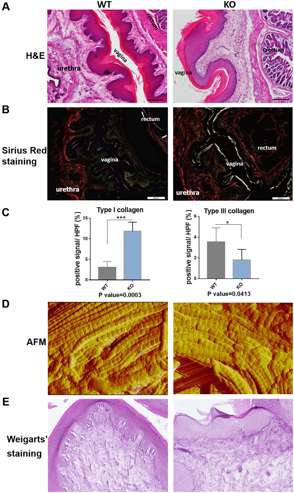
Figure 5.Loxl1 knockout mice reproduce the micro-structural changes of clinical POP. (A) HE staining showed obvious microstructure space and disappeared Vicious Angle structure in Loxl1 knockout mice, scale bar 100 um. (B) Sirius red staining demonstrated increased type I collagen and decreased type III collagen in Loxl1 knockout mice, scale bar 100 um. (C) Sirius red staining as in B was quantified, the proportion of positive signals area (red signals for type I collage and green signals for type III collagen) in each High Power Field of vision (HPF) were quantified, n = 4 in each group. (D) Atom force microscope (AFM) showed aligned collagen fibers in WT mice while disordered arranged collagen fibers in Loxl1 knockout mice. (E) Weigarts’ staining of the vagina tissue showed the elastic fibers in WT mice were linear and polarized, while in the knockout mice were fragmented and arranged disordered.