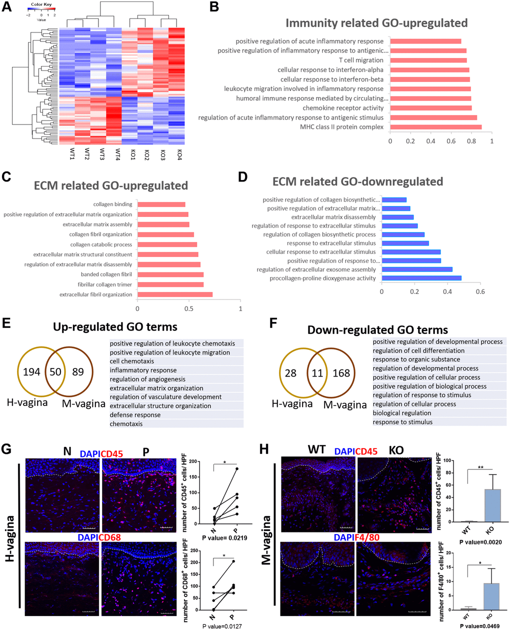
Figure 7.Loxl1 knockout mice recapitulate the molecular changes in vagina tissue of clinical POP. (A) Heat map across all the samples using the top 500 most differently expressed genes in vagina tissue between the Loxl1 knockout mice and WT mice. (B–D) gene ontology categories of differently expressed genes, horizontal axis represent the fold enrichment, p value < 0.05. (E, F) Venn diagram (left) and the form (right) showed the overlaps of up-regulated (E) and down-regulated (F) differentially expressed genes enriched GO terms between human vagina (H-vagina) and mice vagina (M-vagina). (G, H) Immunofluorescence staining for visualization (red) of immune cell infiltration in human vagina (G) and mice vagina (H). The number of positive cells (red) in each High Power Field of vision (HPF) were quantified respectively. (CD45: leukocyte, CD68: macrophage in human, F4/80: macrophage in mice; White dotted line indicated the interface between mucus layer and submucosa layer, n = 5 in each group).