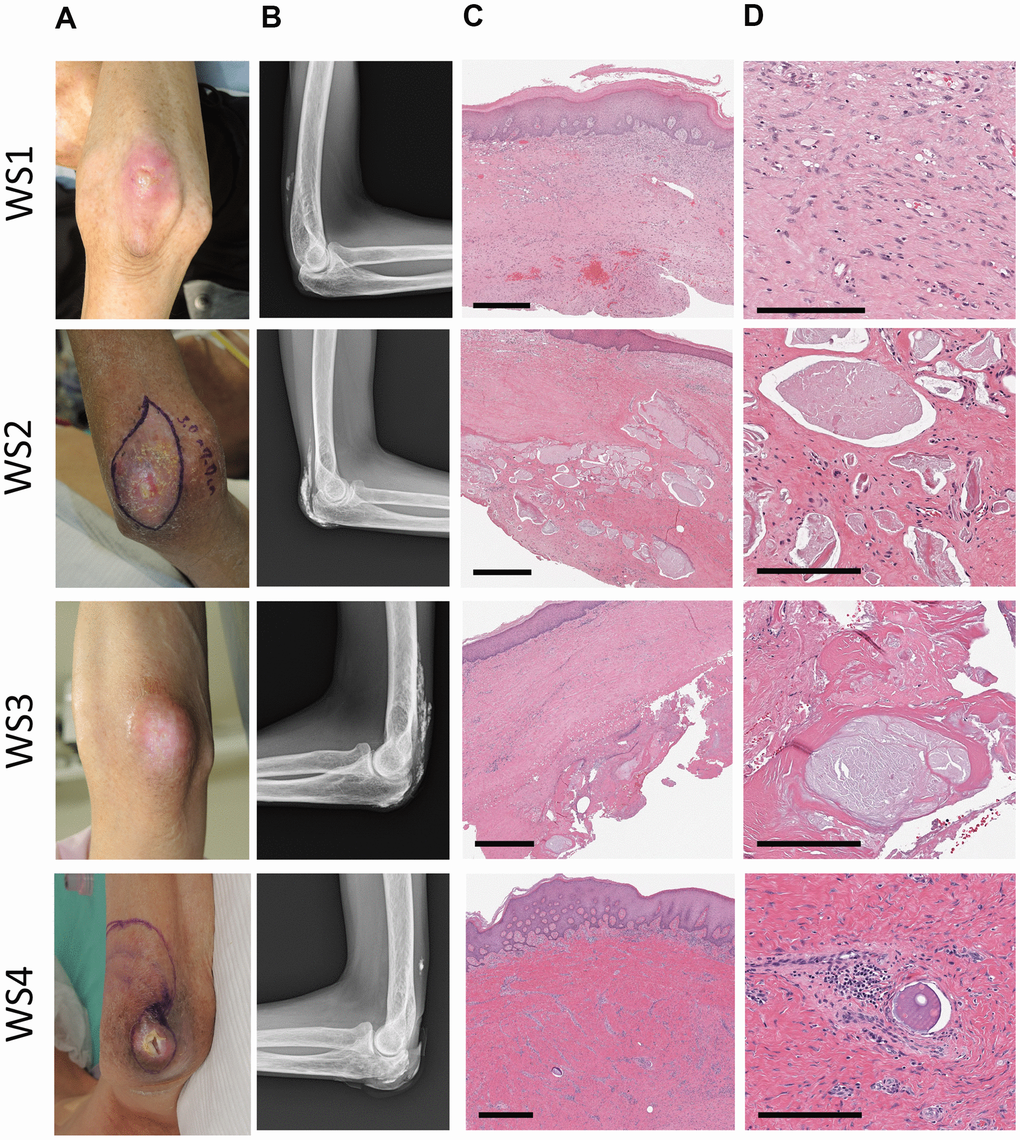
Figure 1.Clinical images of patients. A series of photographs (A) of elbow ulcers in four patients with Werner's syndrome (WS1-4), radiographs (B) of the same area, and HE stained tissue (C, D) of the skin around the ulcers. WS1: 62-year-old male, WS2: 48-year-old male, WS3: 50-year-old female, WS4: 57-year-old male. (A) The skin around the ulcer is sclerotic and atrophic. (B) Various degrees of calcification were observed in the soft tissues around the elbow joint. In WS2 and WS3, it becomes a shadow of the flame state. (C, D) Strong fibrosis of the dermis (all cases) and calcification within the luminal structures (WS2, 3, 4). Scale bars are 700 μm (C) and 200 μm (D).