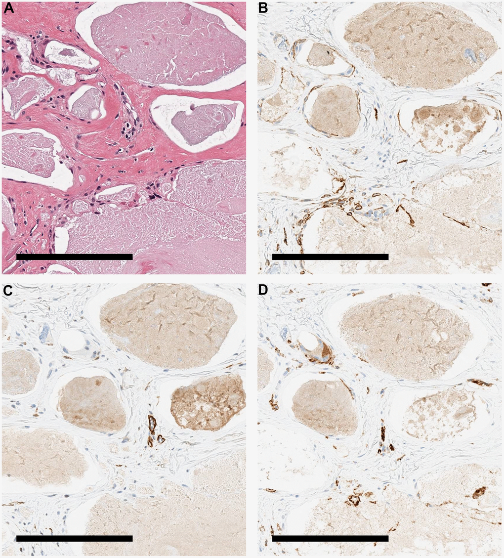
Figure 3.Immunohistochemical staining image of the patient's skin. Various stained tissue images of calcified tissues from a patient with WS. (A) HE. (B) Immunohistochemical staining with podoplanin antibody. Cells around the calcification were positive. (C) Immunohistochemical staining with αSMA antibody. Cells around the calcification were negative. (D) Immunohistochemical staining with CD 31 antibody. The cells around the calcification were weakly positive. All scale bars are 200 μm.