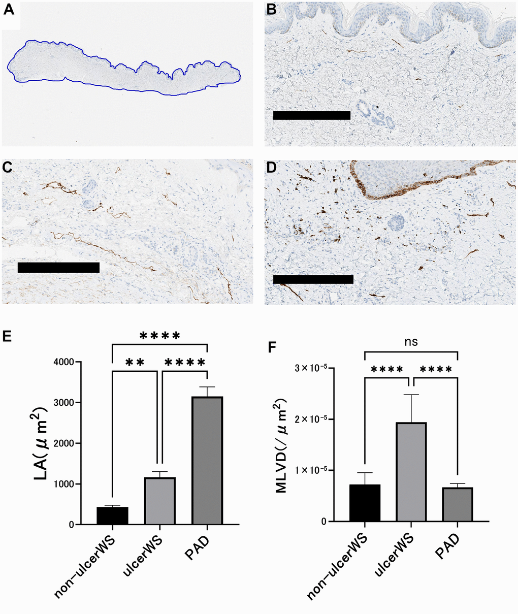
Figure 4.Image analysis of lymphatic vessels in the skin of patients. (A) The area of skin tissue sections was measured by tracing the skin margins using an Aperio Image Scope. (B) A representative example of skin histology taken from an ulcer-free area in a patient with Werner syndrome. Scale bar is 300 μm. The lumen is narrow and small in number. (C) A representative example of skin tissue taken from the ulcer circumference of a patient with PAD. Scale bar is 300 μm. The lumen is dilated and lined up. (D) A representative example of skin tissue taken from the ulcer circumference of a Werner syndrome patient. Scale bar is 300 μm. The lumen is indistinct and randomly proliferating. (E) Comparison of luminal cross-sectional area (LA) of the lymphatic vessels. In four patients with Werner syndrome, two different sites were measured in the non-ulcerated area and in the skin surrounding the ulcer. Three patients with PAD underwent measurements at two different sites of skin around the ulcer. Data are expressed as mean ± standard error. One-way ANOVA followed by Tukey test were performed (* * p < 0.001, **** p < 0.0001). (F) Comparison of lymphatic vessel density (MLVD). In four patients with Werner syndrome, two different sites were measured in the non-ulcerated area and in the ulcer's skin. Three patients with PAD underwent measurements at two different sites of skin around the ulcer. Data are expressed as mean ± standard error. One-way ANOVA followed by Tukey test were performed (n.s. No significant difference, **** p < 0.0001).